
Scalp sarcoidosis is a relatively infrequent disease that mainly affects African-American women.1 To date, very few cases of this disease have been described. We describe 3 patients with scalp sarcoidosis, which manifested as infiltrated plaques with alopecia.
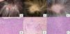
Case 1. The patient was a 50-year-old woman who was admitted to our hospital with exertional dyspnea, photophobia, and skin rash. The physical examination revealed several indurated brown plaques on both lower limbs, as well as partial, irregularly distributed alopecia and scaling erythema (Fig. 1A). Radiography and computed tomography (CT) of the thorax revealed bilateral hilar lymphadenopathy (BHL) and mediastinal lymphadenopathy. Serum levels of angiotensin converting enzyme (ACE) were elevated (72.8 U/L; normal range, 7–25 U/L). The patient had a negative reaction in the tuberculin test. The ophthalmological examination revealed uveitis. Biopsy of the scalp showed non-necrotizing epithelioid granulomas located in the upper to mid dermis with minimal lymphocytic infiltration (Fig. 1B). Naked granulomas were also detected in another sample taken from the leg. The patient was treated with local topical steroids, to which she responded poorly.
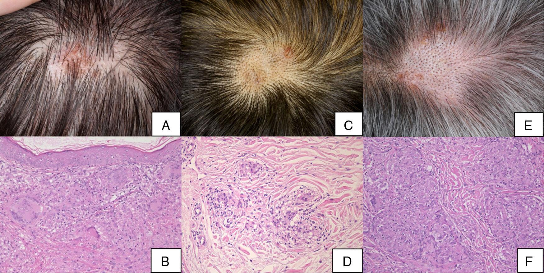
Case 2. The patient was a 51-year-old man who was referred to our department with pulmonary sarcoidosis and uveitis. He complained of blurred vision. The ophthalmological examination revealed nodules in the iris, inflammation of the anterior segment, and vitreous opacity. Radiography and a chest CT scan revealed BHL. Serum ACE levels were elevated (64.5 U/L). The patient had a negative reaction in the tuberculin test. The physical examination revealed indurated reddish-brown plaques and regularly distributed areas of alopecia with black spots on the scalp (Fig. 1C), as well as dark red subcutaneous nodules on the legs. Biopsies of the scalp and legs revealed sarcoid granulomas located in the dermis (Fig. 1D). The patient was treated with local topical steroids, to which he responded poorly.
Case 3. The patient was a 71-year-old man who was referred to our department with uveitis. He reported skin rash on the scalp and the right leg. Physical examination of the scalp revealed multiple erythematous, scaly, indurated areas and an egg-sized, indurated alopecic plaque with black spots (Fig. 1E). There were dark red subcutaneous nodules on the right leg. Radiography and a chest CT scan revealed BHL. Serum ACE levels were elevated (42.5 U/L). The patient had a negative reaction in the tuberculin test. He complained of myodesopsia and the ophthalmological examination revealed iritis and lens opacity. Biopsy of the plaque on the scalp showed sarcoid granulomas extending from the mid to deep dermis (Fig. 1F). The patient was treated with local topical steroids, to which he responded poorly.
All 3 patients were treated with local topical steroids, with a poor outcome in all cases. No systemic treatments were administered.
The clinical presentation of scalp sarcoidosis consists of indurated verrucous plaques and nodular lesions, often accompanied by alopecia, and can resemble that of discoid lupus erythematosus, necrobiosis lipoidica, organoid nevus, and cicatricial alopecia.1–5 In general, scalp sarcoidosis is accompanied by sarcoid skin lesions in other locations.1,2 Our 3 patients had infiltrated plaques with alopecia. Unfortunately, a trichoscopic examination, which is necessary to identify the trichoscopic features of scalp sarcoidosis, was not performed prior to biopsy. The alopecia may have been a consequence of the effects of the sarcoid granuloma on the follicles or of follicular replacement by the granuloma. All 3 patients had cutaneous sarcoidosis on the legs as well as the scalp. Sarcoidosis of other organs is common in patients with scalp sarcoidosis, and pulmonary and ophthalmologic sarcoidosis was observed in all 3 patients. In most cases, scalp sarcoidosis affects patients with active systemic sarcoidosis. We have previous experience with 2 other cases of scalp sarcoidosis in which biopsies were taken from locations other than the scalp. In both cases the patients’ alopecia was strongly suggestive of sarcoidosis. Scalp involvement is common in cases of cutaneous sarcoidosis, and therefore a careful examination of the scalp should be performed if sarcoidosis is suspected. Indeed, scalp sarcoidosis may be less rare than previously thought. It is therefore important to include a scalp examination in the clinical examination of sarcoidosis patients.
Conflicts of InterestThe authors declare that they have no conflicts of interest.
Please cite this article as: Ishikawa M, Ohtsuka M, Yamamoto T. Three Cases of Scalp Sarcoidosis with Alopecia. Actas Dermosifiliogr. 2018;109:933–934.


