A 41-year-old man with no medical or surgical past history of interest was referred to the dermatology service due to skin lesions that had appeared 2 months earlier on both pinnae. The lesions were asymptomatic, and the patient reported no potential triggering event.
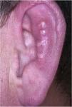
Physical ExaminationPhysical examination revealed 4 pearly papules distributed in a line on the antihelix of the left pinna (Fig. 1) and 2 lesions of similar characteristics on the right pinna.
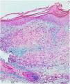
HistopathologyThe histopathology study showed a lymphohistiocytic infiltrate with palisading histiocytes surrounding a central area with necrobiosis and mucin deposit (Fig. 2).
[[?]]What is your Diagnosis?
DiagnosisGranuloma annulare on the antihelix.
Clinical Course and TreatmentIn this case, the lesions resolved after the biopsy and no treatment was required.
CommentGranuloma annulare (GA) is a noninfectious granulomatous skin disease. It is a benign entity of unknown etiology and is common in clinical practice; it especially affects children and young adults and is more frequent in females. It generally presents on the wrists, ankles, hands, and feet in the form of annular erythematous papules with central lightening.1
There are 4 main types of GA. In the local variety, between 80% and 90% of all cases, the lesions appear at the aforementioned sites and with the typical morphology. The generalized form, which represents fewer than 15% of cases, affects the torso and extremities in the form of numerous erythematous or yellowish papules, which may coalesce to form annular or circinate plaques. The subcutaneous variety is rare. It typically affects children and manifests as fast-growing painless subcutaneous nodules, with overlying skin of normal appearance, located on the scalp, pretibial region, buttocks, and palmar and plantar regions. The perforating variety is also rare and manifests as erythematous papules that develop into umbilicated lesions with a squamous or scabbed center. Other less common types of GA have been described and the different types also tend to coincide.2
Very few cases of GA involving the outer ear have been published in the literature.3–6 Although GA is more common in women, most published cases involving the outer ear have been reported in boys or young men,3–6 and a large percentage of these are bilateral.3–5 Moreover, the lesions usually appear on the most exposed areas and it has therefore been suggested that small, repeated traumas may be implicated as a trigger, even when patients do not always identify them.
We report an atypical GA in terms of site of the lesions and in terms of morphology. This presentation is rare in the literature and a correct diagnosis, which is essential to avoid unnecessary interventions or treatments, with their potential complications, may pose a challenge.
Conflicts of interestThe authors declare that they have no conflicts of interest.
The authors would like to thank the Dermatology department of Complejo Hospitalario Universitario de Pontevedra, Spain, for their cooperation.
Please cite this article as: Oro-Ayude M, Hernández Cancela RM, Flórez A. Pápulas perladas en el antihélix. Actas Dermosifiliogr. 2020;111:603–604.


