


Pseudomonas aeruginosa causes multiple cutaneous infections, which are typically mild in immunocompetent individuals but can be potentially serious in immunocompromised patients. These include interdigital intertrigo, green foot syndrome, folliculitis (swimming pool or hot-tub folliculitis), pyodermatitis vegetans, balanitis, otitis externa (swimmer's otitis), malignant otitis externa, omphalitis of newborn, ecthyma gangrenosum, cellulitis, abscesses, nodules, necrotizing fasciitis, and superinfection of burn wounds, surgical wounds, and diabetic, decubitus, or venous foot ulcers.1
Green nail, also known as chloronychia or green nail syndrome, is usually caused by infection with P aeruginosa. The clinical presentation consists of a classic triad of green discoloration (yellowish green, brownish-green, bluish-green, or black-green) of the ungueal lamina, proximal chronic paronychia, and distolateral onycholysis. Most strains of this bacterium produce pigments, including the yellowish-green fluorescein pyoverdine and blackish-green pyocyanin (1-hydroxy-5-methyl-phenazine), which give rise to the typical green color of infected nails. The differential diagnosis includes subungual hematoma, malignant melanoma, jaundice, blistering disease, yellow nail syndrome, infections caused by other agents such as Trichosporon inkin and Aspergillus, Candida, and Proteus species, drug-induced nail discoloration, and exogenous pigmentation caused by chemical substances.2,3
Factors that predispose individuals to green nail due to P aeruginosa infection include immunocompromise, diabetes mellitus, nail trauma, damp environments, and prolonged exposure to water, soap, or detergents. Nails previously affected by onycholysis, paronychia, onychotillomania, or nail psoriasis are more vulnerable.2 A strong relationship between fungal and P aeruginosa nail infections has also been reported.4,5
We present the clinical and dermoscopic findings of an immunocompetent male patient with green nail caused by onychomycosis (OM) coinfected with P aeruginosa.
The patient was a 39-year-old man with no personal history of interest who was seen for discoloration of a toenail on his right foot that had begun 2 years earlier. He reported no previous trauma. He had been treated with a 5% amorolfine solution twice per week for 3 months, without improvement.
Physical examination revealed subungual hyperkeratosis and yellow-green discoloration of the nail plate of the fifth toe of the right foot (Fig. 1A and B). Dermoscopy revealed alterations of the nail, subungual hyperkeratosis, an irregular distal border, and a multicolored pattern on the nail plate consisting of areas of homogeneous greenish and blackish-blue coloration and other longitudinally distributed yellow-colored areas (Fig. 1C). The interdigital spaces of both feet were macerated and fissured.
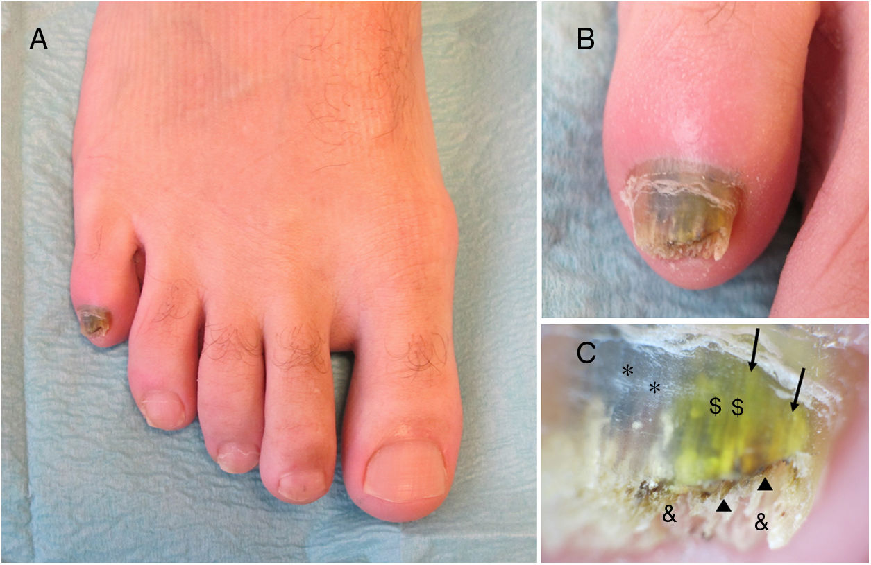
Nail of the fifth toe of the right foot. A, Yellow-green discoloration of the nail plate. B, Subungual hyperkeratosis and areas of yellow, green, and black discoloration. C, Dermoscopic image showing subungual hyperkeratosis (&), an irregular distal border, brown areas with a hemorrhagic appearance (⏶), and discoloration of the nail plate with homogeneous areas of green ($) and blackish-blue (*) and longitudinally distributed areas of yellowish discoloration (→).
The suspected diagnosis was tinea pedis associated with OM or P aeruginosa nail infection. Several samples of the affected nail were collected for fungal and bacterial culture. Culture revealed growth of P aeruginosa and a filamentous fungus that could not be identified due to bacterial overgrowth. Initial treatment, selected based on the results of an antibiogram, consisted of oral ciprofloxacin (500 mg/12 h) for 10 days, and was followed by oral terbinafine (250 mg/d) for 3 months. Follow-up evaluation after 6 months revealed complete resolution of the infectious process.
Tinea pedis (athlete's foot) is considered the most prevalent dermatophytic infection and most often affects the interdigital spaces. The simple form is asymptomatic or mildly pruritic and is characterized by erythema, scaling, and cracking. The third and fourth interdigital spaces are most commonly affected. Some nondermatophyte fungi and bacteria (Corynebacterium minutissimum) cause similar clinical manifestations. The complex form of tinea pedis interdigitalis is characterized by secondary bacterial superinfection and a more severe presentation. It usually causes itching or pain with inflammation, maceration, erosions, and foul odor.6,7
OM is an infection of the nails caused by dermatophyte fungi (tinea unguium), nondermatophyte filamentous fungi, or yeast. The presumptive diagnosis is established based on clinical features and should be confirmed by mycological analyses (direct examination and culture) or histology. Dermoscopy (onychoscopy) can be diagnostically useful. Depending on the clinical presentation, OM is classified as distal and lateral subungual, superficial (black and white), proximal subungual, endonyx, mixed pattern, total dystrophic, or secondary.5
P aeruginosa coinfection has been described in patients with OM,4,5 as well as those with tinea pedis.7 In these cases there is subungual hyperkeratosis and yellow-green discoloration of the affected nails.4 The presence of fungi favors colonization and promotes the growth of P aeruginosa. In cases of coinfection with P aeruginosa isolation of the causative fungus may be impossible due to bacterial overgrowth in culture or the bacteria's fungistatic and/or fungicidal properties.4,7 In fact, this bacterium produces substances that inhibit in vitro growth of yeasts (Candida albicans), nondermatophyte filamentous fungi (Aspergillus fumigatus and Fusariumsolani), and dermatophyte fungi (Trichophyton mentagrophytes and Trichophyton rubrum).8,9
In the present case culture of a nail sample allowed isolation, but not identification, of a filamentous fungus. We were unable to determine whether the fungus in question was dermatophytic or not. Moreover, we did not collect samples from the interdigital spaces of the patient. The growth of dermatophytes such as Trichopyton species in cultures of these samples could have confirmed a clinical diagnosis of tinea pedis. Because the clinical, dermoscopic, and microbiological findings were compatible with a mixed bacterial (P aeruginosa) and fungal nail infection, we decided to prescribe treatment with ciprofloxacin and an oral antifungal for 3 months. Another useful therapeutic option in cases in which a single nail is affected is photodynamic therapy, which can be combined with concurrent tinea pedis treatment.10
In conclusion, we describe a new case of green nail caused by OM coinfected with P aeruginosa in an immunocompetent adult male. Adequate treatment is essential in cases of OM with concomitant fungal and Pseudomonas infection. Because the interaction between the 2 agents can complicate microbiological diagnosis, it is important to be familiar with their respective clinical and dermoscopic features.
Conflicts of InterestThe authors declare that they have no conflicts of interest.
Please cite this article as: Monteagudo B, Figueroa O, Suárez-Magdalena O, Méndez-Lage S. Uña verde causada por onicomicosis coinfectada por Pseudomonas aeruginosa Eczema y urticaria en Portugal. 2019;110:783–785.


