Since the emergence of COVID-19 infection in Spain, in the beginning of February 2020, the most populated regions, such as Madrid and Catalonia, have concentrated the highest case incidence. Suspect of COVID-19 is based on clinical signs comprising fever, cough, fatigue, ageusia, anosmia, myalgia and dyspnea. Laboratory alterations include lymphopenia, increased LDH, D Dimer, Ferritin and CRP. The diagnosis is based on virus detection in oropharyngeal swabs. Some rapid tests are available for seroconversion detection, for determination of specific IgM and IgG, but its use is still limited due to low availability, and uncompleted validation. Restricted measures have been performed in the Spanish population. Alarm state and general confinement in Spain started in March 14th and has not yet been finished.
A number of dermatoses associated with covid-19 infection have already been described,1 including erythematous or purpuric rash in the trunk, urticarial and chickenpox-like lesions. Acro-ischemic lesions have been reported in intensive care unit-admitted patients,2 due to disseminated intravascular coagulation (DIC) and clinically behaving as a dry gangrene. A case of acute acro-ischemic lesions in a child has recently been reported in Italy.3
During the first weeks of April, national and regional social media have reported a vast number of chilblain-like purpuric acral lesions in hands and feet, predominating in children, and up to date, accurate descriptions in the existing medical literature have been sparse.3 In Spain, reports of these lesions have been widespread in the territory, but they have been more frequently reported on the most populated areas, the Metropolitan Conurbations of Madrid and Barcelona.
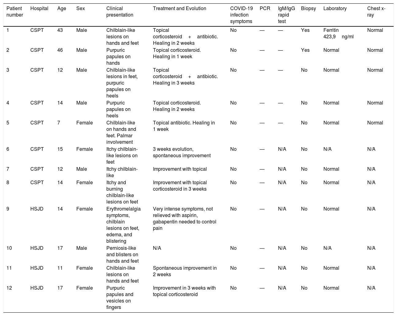
We have retrospectively reviewed 12 cases of acral purpuric lesions that have been studied thoroughly in two hospitals in the Barcelona area (Consorci Sanitari Parc Taulí and Hospital Sant Joan de Deu). Their clinical characteristics are shown in Table 1. None of them had COVID-related clinical manifestations. Most patients were children and young adults. Clinical picture comprised two types of lesions: 1. Acral erythematous purpuric lesions in fingers and toes, with accompanying edema, similar to common chilblains or pernio (Figure 1), sometimes evolving into blisters, and crusts. Hands and feet were not cold, and the symptoms usually ranged from itch to burning or painful sensations; 2. Papular or macular purpuric round-shaped lesions, 5 to 8mm of diameter, in palmar or plantar surfaces, or over the heels (Figure 2), clinically resembling vasculitis or erythema multiforme, usually asymptomatic or itchy. The two types of lesions were present alone or in combination.
Patients’ characteristics.
| Patient number | Hospital | Age | Sex | Clinical presentation | Treatment and Evolution | COVID-19 infection symptoms | PCR | IgM/IgG rapid test | Biopsy | Laboratory | Chest x-ray |
|---|---|---|---|---|---|---|---|---|---|---|---|
| 1 | CSPT | 43 | Male | Chilblain-like lesions on hands and feet | Topical corticosteroid+antibiotic. Healing in 2 weeks | No | — | — | Yes | Ferritin 423,9ng/ml | Normal |
| 2 | CSPT | 46 | Male | Purpuric papules on hands | Topical corticosteroid. Healing in 1 week | No | — | — | Yes | Normal | Normal |
| 3 | CSPT | 12 | Male | Chilblain-like lesions in feet, purpuric papules on heels | Topical corticosteroid+antibiotic. Healing in 3 weeks | No | — | — | No | Normal | Normal |
| 4 | CSPT | 14 | Male | Purpuric papules on heels | Topical corticosteroid. Healing in 2 weeks | No | — | — | No | Normal | Normal |
| 5 | CSPT | 7 | Female | Chilblain-like on hands and feet. Palmar involvement | Topical antibiotic. Healing in 1 week | No | — | — | No | Normal | Normal |
| 6 | CSPT | 15 | Female | Itchy chilblain-like lesions on feet | 3 weeks evolution, spontaneous improvement | No | — | N/A | No | N/A | N/A |
| 7 | CSPT | 12 | Male | Itchy chilblain-like | Improvement with topical | No | — | N/A | No | Normal | N/A |
| 8 | CSPT | 14 | Female | Itchy and burning chilblain-like lesions on feet | Improvement with topical corticosteroid in 3 weeks | No | — | N/A | No | Normal | N/A |
| 9 | HSJD | 14 | Female | Erythromelalgia symptoms, chilblain lesions on feet, edema, and blistering | Very intense symptoms, not relieved with aspirin, gabapentin needed to control pain | No | — | N/A | No | Normal | N/A |
| 10 | HSJD | 17 | Male | Perniosis-like and blisters on hands and feet | N/A | No | — | N/A | No | N/A | N/A |
| 11 | HSJD | 11 | Female | Chilblain-like lesions on hands and feet | Spontaneous improvement in 2 weeks | No | — | N/A | No | Normal | N/A |
| 12 | HSJD | 17 | Female | Purpuric papules and vesicles on fingers | Improvement in 3 weeks with topical corticosteroid | No | — | N/A | No | Normal | N/A |
CSPT: Consorci Sanitari Parc Taulí; HSJD: Hospital Sant Joan de Deu; N/A: not available.
The results of COVID-19 screening yielded negative results in all cases, including specific PCR and a rapid test for IgM/IgG antibodies (VivaDiag, VivaCheck Biotech, Hangzhou, China), performed only in 5 patients due to current low availability. Laboratory tests (including blood cell count, ferritin, CRP, D Dimer, Ferritin and LDH) and chest X-ray, when available, showed normal or negative results. In two patients, a histopathological study of a punch biopsy of the lesions yielded nonspecific findings, with dermal edema, sparse keratinocyte necrosis, and a deep mixed infiltrate with mostly perivascular or perieccrine reinforcement.
All cases showed a good evolution, achieving a complete healing after two to three weeks of topical corticosteroid or combination of topical corticosteroid plus topical antibiotic. In only one patient, pain control needed the administration of oral gabapentin. None of the patients have develop any COVID-related clinical manifestation since the diagnosis of skin lesions.
As a conclusion, this stunning clinical picture is atypical because it has appeared during warm weather, not associated with common chilblains, and although it has a temporal relationship with COVID pandemic, in none of the patients an evidence of current or past COVID-19 infection could be demonstrated. Epidemiological association is clear with COVID-19 pandemic, but our results show that such cutaneous lesions are not a manifestation of active coronavirus infection, as determined by currently available tests. One explanation could be an early contact with COVID-19 during the month of February or early March, without clinical symptoms, which would have made the virus undetectable with PCR technique. A second explanation could be low sensibility of the rapid IgG/IgM tests, or a fast disappearance of circulating antibodies, with low levels that do not match the technique detection threshold. A third one would comprise different etiopathogenic factors related to confinement, which have not been identified up to date. A more detailed study of more skin biopsies, self-immunity profiling in serum, and more refined quantitative PCR detection are in course, in order to elucidate the cause of these dermatological lesions.
Conflict of interestThe authors declare that they have no conflict of interest.
Please cite this article as: Romaní J, Baselga E, Mitjà O, Riera-Martí N, Garbayo P, Vicente A, et al. Lesiones pernióticas y acrales en España durante el confinamiento por COVID: análisis retrospectivo de 12 casos. Actas Dermosifiliogr. 2020;111:426–429.


