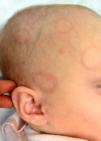
An 8-month-old infant girl with no significant personal or obstetric history was taken to the emergency department for evaluation of lesions on the face and scalp that had appeared 2 weeks earlier. The patient had had no fever at any time and no other associated symptoms. The only history of interest was an upper airway cough the week before the lesions appeared, which did not require treatment. The mother and other close relatives mentioned no previous skin disorders or medical conditions of interest.
Physical ExaminationPhysical examination revealed several asymptomatic, erythematous, edematous, nonscaly annular plaques in a symmetrical distribution (Figs. 1 and 2). The physical examination was otherwise normal.
Additional TestsThe results of the blood workup—including tests for antinuclear antibodies, anti-Ro antibodies, anti-La antibodies, and anti-ribonucleoprotein antibodies; serologies; and hormonal profile—were negative or normal in the mother and the patient.
Clinical Course and TreatmentAs there were no signs of severity or specific disease, the condition was managed with observation. The lesions remitted spontaneously 2 weeks later, leaving no sequelae, and did not recur in the following 6 months.
What Is Your Diagnosis?
DiagnosisGiven the characteristic clinical presentation, the favorable clinical course, and the spontaneous resolution of the lesions, we established a clinical diagnosis of annular erythema of infancy, probably a reactive process in the context of the earlier upper airway infection.
CommentThe figurate erythemas are a group of dermatoses in which the primary lesion has an annular, arciform, polycyclic, or concentric morphology. Many dermatologic diseases can adopt annular forms (urticaria, erythema multiforme, tinea corporis,1 granuloma annulare, pityriasis rosea, sarcoidosis, lupus erythematosus, etc.). However, there are also entities characterized by circinate or annular lesions that can be fixed or migratory and whose appearance is related to drug hypersensitivity, infections, neoplastic disease, insect bites, or autologous hypersensitivity.2,3
Many classifications of figurate erythemas have been published in the medical literature. These eruptions can be classified according to the predominant cellular component identified by histologic study (lymphocytes, neutrophils, eosinophils, granulomas, or plasma cells), by the clinical characteristics of the primary lesion (macular, urticarial, desquamative, or raised), or by whether the etiology is known or unknown.2,4–6
Annular erythema of infancy is a rare, benign form of figurate erythema of unknown etiology. It typically appears in the first months of life in the form of erythematous, annular, slow-growing papules with raised borders and a central clearing. The lesions can appear on the face and trunk and usually resolve after a few days without sequelae. New lesions may appear and follow the same course. Histology shows both superficial and deep perivascular lymphohistiocytic infiltrates with abundant eosinophils. In a variant called neutrophilic figurate erythema of infancy, histology also shows leukocytoclasia. In this variant, similar lesions initially manifest on the face and resolve spontaneously after 2 to 4 weeks. The condition tends to be chronic and the lesions can reappear on the limbs.4,5
In conclusion, figurate erythemas are not specific entities but rather reaction patterns that can vary from person to person. Because of the similarities in clinical presentation, these conditions are difficult to diagnose and in some cases can only be distinguished by subtle differences in clinical or histologic features. In managing these conditions, it is important to rule out diseases that require specific treatment or endanger the health of the patient. In cases highly suggestive of annular erythema of infancy, given the benign nature of the lesions, immediate biopsy is not necessary, and watchful waiting with periodic follow-up is appropriate.4,6
Conflicts of InterestThe authors declare that they have no conflict of interest.
Please cite this article as: Sánchez-Orta A, Albízuri-Prado F, Feito-Rodriguez M. Lesiones anulares en cabeza de un lactante. Actas Dermosifiliogr. 2016;107:338–339.


