Facial reconstruction surgery requires detailed knowledge of anatomic and functional structures such as the nose, eyelids, and lips, because of the importance of preserving their function, shape, and cosmetic appearance.
Various surgical techniques are available to repair defects; the quickest and simplest reconstruction is by direct closure. First, an ellipse must be created; this involves lengthening the surgical incision to eliminate the excess skin at each end of the incision. One technique used to remove excess skin is the Burow triangle or wedge-shaped resection.1
The lower eyelid is formed by the orbicularis muscle covered by thin and lax skin. The infraorbital region is the area immediately inferior and medial to the lower eyelid, and the malar region is inferior and lateral to this eyelid; lower down these 2 regions give rise to the cheek. These facial cosmetic areas have different textures, colors, and densities, from a fine skin with minimal subcutaneous cellular tissue in the eyelid to the thicker skin of the cheek, strongly adherent to the subcutaneous cellular tissue.2
In the repair of defects that affect the infraorbital or malar regions, the skin of the cheek is united with the skin of the eyelid, despite these marked differences, and there is the associated risk of provoking eversion of the palpebral margin, separating it from the surface of the eye and producing ectropion.3
We present a simple option for closure that minimizes this possibility.
Surgical TechniqueFirst Step: DesignThe elliptical excision must be marked before performing anesthesia because the anesthetic injection will distort the anatomy. A Burow triangle is then designed at the medial end (Fig. 1A).
A, Design of the closure; the Burow triangle is designed at the medial end of the incision. B, Anchorage of the ellipse; a to a’ orientation of the first stitch. C, Diagonal movement of the skin. D, Final closure. The points indicate the area of greatest tension when the borders are united.
It is important to dissect the tissues in the direction of the cheek to achieve better displacement. Do not dissect towards the eyelid because of the fragility of the skin in that area.
Third Step: AnchorageThe first stitch must be placed at the point of greatest tension, which is between points a and a’ shown in figure 1 B, C, and D. This is performed with a nonabsorbable intradermal suture.
Fourth Step: External SuturesWe prefer to use horizontal U stitches buried in the lower eyelid.
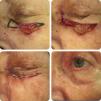
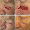
Figures 2 and 3 show patients in whom malignant tumors were excised. Closure of the defect was performed using the technique described.
- 1.
It is essential to make the first stitch a to a’ in a diagonal orientation (Fig. 1B) to prevent tension perpendicular to free border of the eyelid, which would provoke ectropion.
- 2.
Protect the skin of the lower eyelid, as it is thin and delicate.
- 3.
The intradermal suture can cause ectropion if fibers of the orbicularis muscle are included in the suture.3
We have proposed this technique for infraorbital defects of up to 1cm diameter. In a previous article we described a rotation-advancement flap for larger defects.4
The use of the Burow triangle in the ellipse is not only to remove excess skin, but also to move the point of greatest tension, transferring it to the medial sector of the eyelid, which will minimize the risk of ectropion (Fig. 1, C and D).
Please cite this article as: Molinari LM, Ferrario D, Galimberti GN. Use of the Burow Triangle or Wedge-shaped Resection During the Repair of Infraorbital Defects. Actas Dermosifiliogr. 2015;106:689–691.


