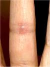
A 40-year-old woman presented with sudden onset of a painful nodule on the palmar aspect of the left third digit, non-dominant hand. The nodule had been present for 4 days. No previous local trauma or Raynaud's phenomenon were found in the past history, which was also unremarkable for smoking, drug intake or other comorbidities.
Physical ExaminationA bluish-violet nodule of 4 mm was observed on the palmar side of the third digit, at the level of the proximal interphalangeal (PIP) joint (Fig.1). The nodule was highly sensitive on compression and it was fixed to the subcutaneous structures. No color change on the tip of the digit was present and pulsation at both the left radial and ulnar arteries was normal. Physical examination was not otherwise remarkable.
Complementary TestsDermoscopic examination of the lesion showed a blue structureless pattern (homogeneous blue pattern) (Fig. 2).
An excisional biopsy under intrathecal anesthesia was performed.
Histological analysis revealed a severely ectatic venous vascular structure with intraluminal congestion and an organized thrombus in its interior. A scarce surrounding inflammatory infiltrate containing lymphocytes and occasional neutrophils was also observed (Fig. 3).
Laboratory studies including complete blood count, prothrombin time, activated partial thromboplastin time, proteins C and S, factor V Leiden, antithrombin III, anticardiolipin antibodies and lupus anticoagulant did not showed relevant data.
DiagnosisBased on the clinical and histological findings, the patient was diagnosed with thrombosis of palmar digital vein.
Course and TreatmentAfter surgical excision symptoms completely resolved and recurrence of the lesion did not occur.
CommentsThrombosis of the palmar digital veins was first described in 1936 by Jadassohn.1 Given the scarcity of cases reported in the literature digital vein thrombosis seems to be a relatively rare condition.2
It is observed most often in females between 35 and 65 years although it can occur at any age.3 Typically it presents as one or more hard painful nodules on the palmar or lateral side of the fingers, with a bluish-black or erythematous discoloration. The fourth digit is most often affected, sometimes but not always associated with local trauma like wearing a ring.3 Typically, as in our patient, the nodule is located around the PIP joint, but nodules have also been located over the middle or proximal phalanx or at the level of the distal interphalangeal joint.
Although the etiology is still to be completely understood several factors have been implicated, namely the uniqueness of the anatomic characteristics of the palmar digital veins. Different from dorsal veins, these vessels are surrounded by a sheath of fine connective tissue and fat cushions.4 If this supporting structure deteriorates through aging these veins become more prone to collapse leading to thrombosis. Furthermore, because of their localization at the flexion creases of the fingers and the fact that palmar veins are smaller in diameter and contain more valves than dorsal veins, these seems to be more vulnerable to trivial trauma.5 Such injuries are presumably unrecognized and repetitive. As the majority of the cases our patient could not recall a directly preceding traumatic event. However, it should be noted that she is a dermatologic surgeon. Therefore, minor trauma may have contributed.
It has also been suggested that hypercoagulable states may play a role. However an association between digital palmar vein thrombosis and hemostatic abnormalities has been difficult to prove.3,6 Nevertheless, evaluation of the hemostatic profile may be considered but has not been proved to contribute to either the diagnosis or the management.
The differential diagnosis should include varices with no thrombosis, hemangiomas, epidermal inclusion cysts, giant-cell tumors, ganglions, lipomas, blue nevi, and melanoma metastasis.6
Conservative management such as massaging the lesion, applying hot compresses or compression bandages and taking aspirin seems to be effective in relieving of symptoms.3 Surgical excision is indicated when the clinical diagnosis is in doubt or if symptoms persist.
This case highlights the importance to consider palmar digital vein thrombosis in the differential diagnosis of a painful, firm, blue nodule located at or in close proximity to one of the flexion crease of a finger.
Conflicts of InterestThe authors declare no conflicts of interest.
Please cite this article as: Batista M, Brinca A, Vieira R. Aparición súbita de un nódulo azulado doloroso en la mano. Actas Dermosifiliogr. 2021;112:843–844.


