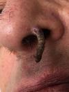
A 65-year-old man presented in our Dermatology department with a solitary hyperkeratotic filiform papule with exophytic growth and an infiltrated base on the left nasal vestibule with few months of evolution (Fig. 1).
Complete excision of the lesion was performed and histopathology revealed an epidermal lesion of endophytic growth formed by mature keratinocytes in the central axis and basaloid cells in the periphery (Fig. 2). There were also intralesional corneal cysts surrounded by a dermal inflammatory infiltrate of lymphocytic predominance. These findings confirmed the diagnosis of inverted follicular keratosis (IFK).
IFK is a rare benign tumor originating from the infundibular portion of the hair follicle with an exoendophytic growth. It usually presents as an asymptomatic solitary verrucous facial papule, more common in elderly males.
A broad spectrum of differential diagnosis is possible, such as seborrheic keratosis, viral warts, squamous-cell carcinoma, keratoacanthoma or basal cell carcinoma. Clinicopathologic correlation is crucial since the diagnosis is generally made by histology. Instead, some authors consider this entity a rare variant of seborrheic keratosis.
Complete surgical excision is the best treatment option to avoid recurrences.
This curious lesion is well-recognized by most dermatopathologists but sometimes underdiagnosed by dermatologists making it an interesting diagnostic challenge.
We are indebted to Ana Marques, MD, from the Department of Pathology in Centro Hospitalar São João EPE, Porto, for her contribution to the histological diagnosis of the lesion as well as her willingness to collaborate in this work.


