array:25 [
  "pii" => "S1578219021001438"
  "issn" => "15782190"
  "doi" => "10.1016/j.adengl.2021.03.007"
  "estado" => "S300"
  "fechaPublicacion" => "2021-06-01"
  "aid" => "2539"
  "copyright" => "AEDV"
  "copyrightAnyo" => "2020"
  "documento" => "simple-article"
  "crossmark" => 1
  "licencia" => "http://creativecommons.org/licenses/by-nc-nd/4.0/"
  "subdocumento" => "crp"
  "cita" => "Actas Dermosifiliogr. 2021;112:558-9"
  "abierto" => array:3 [
    "ES" => true
    "ES2" => true
    "LATM" => true
  ]
  "gratuito" => true
  "lecturas" => array:1 [
    "total" => 0
  ]
  "Traduccion" => array:1 [
    "es" => array:20 [
      "pii" => "S0001731020305044"
      "issn" => "00017310"
      "doi" => "10.1016/j.ad.2019.10.010"
      "estado" => "S300"
      "fechaPublicacion" => "2021-06-01"
      "aid" => "2539"
      "copyright" => "AEDV"
      "documento" => "simple-article"
      "crossmark" => 1
      "licencia" => "http://creativecommons.org/licenses/by-nc-nd/4.0/"
      "subdocumento" => "crp"
      "cita" => "Actas Dermosifiliogr. 2021;112:558-9"
      "abierto" => array:3 [
        "ES" => true
        "ES2" => true
        "LATM" => true
      ]
      "gratuito" => true
      "lecturas" => array:1 [
        "total" => 0
      ]
      "es" => array:11 [
        "idiomaDefecto" => true
        "cabecera" => "<span class="elsevierStyleTextfn">Carta cient&#237;fico-cl&#237;nica</span>"
        "titulo" => "Colgajo de Antia-Buch para un defecto amplio del polo superior de la oreja"
        "tienePdf" => "es"
        "tieneTextoCompleto" => "es"
        "paginas" => array:1 [
          0 => array:2 [
            "paginaInicial" => "558"
            "paginaFinal" => "559"
          ]
        ]
        "titulosAlternativos" => array:1 [
          "en" => array:1 [
            "titulo" => "Antia-Buch Flap for a Large Upper-Pole Defect of the Ear"
          ]
        ]
        "contieneTextoCompleto" => array:1 [
          "es" => true
        ]
        "contienePdf" => array:1 [
          "es" => true
        ]
        "resumenGrafico" => array:2 [
          "original" => 0
          "multimedia" => array:7 [
            "identificador" => "fig0015"
            "etiqueta" => "Figura 3"
            "tipo" => "MULTIMEDIAFIGURA"
            "mostrarFloat" => true
            "mostrarDisplay" => false
            "figura" => array:1 [
              0 => array:4 [
                "imagen" => "gr3.jpeg"
                "Alto" => 835
                "Ancho" => 905
                "Tamanyo" => 98514
              ]
            ]
            "descripcion" => array:1 [
              "es" => "<p id="spar0015" class="elsevierStyleSimplePara elsevierViewall">Esquema paso a paso de la t&#233;cnica de Antia-Buch para el polo superior&#58; lesi&#243;n en el polo superior de la oreja &#40;a&#41;&#46; Defecto despu&#233;s de la resecci&#243;n del tumor &#40;b&#41;&#46; Dise&#241;o de los colgajos &#40;c&#41;&#46; Disecci&#243;n de los colgajos condrocut&#225;neos &#40;d&#41;&#46; Posicionamiento y sutura de los colgajos &#40;e&#41;&#46;</p>"
            ]
          ]
        ]
        "autores" => array:1 [
          0 => array:2 [
            "autoresLista" => "J&#46;C&#46; Barrera Gamboa, A&#46;E&#46; Acosta Madiedo de Hart"
            "autores" => array:2 [
              0 => array:2 [
                "nombre" => "J&#46;C&#46;"
                "apellidos" => "Barrera Gamboa"
              ]
              1 => array:2 [
                "nombre" => "A&#46;E&#46;"
                "apellidos" => "Acosta Madiedo de Hart"
              ]
            ]
          ]
        ]
      ]
      "idiomaDefecto" => "es"
      "Traduccion" => array:1 [
        "en" => array:9 [
          "pii" => "S1578219021001438"
          "doi" => "10.1016/j.adengl.2021.03.007"
          "estado" => "S300"
          "subdocumento" => ""
          "abierto" => array:3 [
            "ES" => true
            "ES2" => true
            "LATM" => true
          ]
          "gratuito" => true
          "lecturas" => array:1 [
            "total" => 0
          ]
          "idiomaDefecto" => "en"
          "EPUB" => "https://multimedia.elsevier.es/PublicationsMultimediaV1/item/epub/S1578219021001438?idApp=UINPBA000044"
        ]
      ]
      "EPUB" => "https://multimedia.elsevier.es/PublicationsMultimediaV1/item/epub/S0001731020305044?idApp=UINPBA000044"
      "url" => "/00017310/0000011200000006/v1_202106081644/S0001731020305044/v1_202106081644/es/main.assets"
    ]
  ]
  "itemSiguiente" => array:20 [
    "pii" => "S1578219021000688"
    "issn" => "15782190"
    "doi" => "10.1016/j.adengl.2021.02.002"
    "estado" => "S300"
    "fechaPublicacion" => "2021-06-01"
    "aid" => "2583"
    "copyright" => "AEDV"
    "documento" => "simple-article"
    "crossmark" => 1
    "licencia" => "http://creativecommons.org/licenses/by-nc-nd/4.0/"
    "subdocumento" => "crp"
    "cita" => "Actas Dermosifiliogr. 2021;112:559-63"
    "abierto" => array:3 [
      "ES" => true
      "ES2" => true
      "LATM" => true
    ]
    "gratuito" => true
    "lecturas" => array:1 [
      "total" => 0
    ]
    "en" => array:11 [
      "idiomaDefecto" => true
      "cabecera" => "<span class="elsevierStyleTextfn">Case and Research Letters</span>"
      "titulo" => "Ultrasound Features of Facial and Extrafacial Granuloma Facial&#46; A Caso Series"
      "tienePdf" => "en"
      "tieneTextoCompleto" => "en"
      "paginas" => array:1 [
        0 => array:2 [
          "paginaInicial" => "559"
          "paginaFinal" => "563"
        ]
      ]
      "titulosAlternativos" => array:1 [
        "es" => array:1 [
          "titulo" => "Caracter&#237;sticas ecogr&#225;ficas del granuloma facial y extrafacial&#46; Una serie de casos"
        ]
      ]
      "contieneTextoCompleto" => array:1 [
        "en" => true
      ]
      "contienePdf" => array:1 [
        "en" => true
      ]
      "resumenGrafico" => array:2 [
        "original" => 0
        "multimedia" => array:8 [
          "identificador" => "fig0005"
          "etiqueta" => "Figure 1"
          "tipo" => "MULTIMEDIAFIGURA"
          "mostrarFloat" => true
          "mostrarDisplay" => false
          "figura" => array:1 [
            0 => array:4 [
              "imagen" => "gr1.jpeg"
              "Alto" => 2803
              "Ancho" => 1417
              "Tamanyo" => 405826
            ]
          ]
          "detalles" => array:1 [
            0 => array:3 [
              "identificador" => "at0005"
              "detalle" => "Figure "
              "rol" => "short"
            ]
          ]
          "descripcion" => array:1 [
            "en" => "<p id="spar0005" class="elsevierStyleSimplePara elsevierViewall">Granuloma faciale&#46; A&#46; Case 1&#58; erythematous-violaceous plaque on the right cheek&#46; B&#46; Hypoechoic&#44; heterogenous lesion in dermis and hypodermis&#44; measuring 4&#44;3&#8239;mm thick&#46; Note the subepidermal hypoechoic band &#40;22&#8239;MHz probe&#44; B-mode&#41;&#46; C&#46; Increased vascularity on color Doppler mode&#46;</p>"
          ]
        ]
      ]
      "autores" => array:1 [
        0 => array:2 [
          "autoresLista" => "D&#46; Morgado-Carrasco, P&#46; Giavedoni, J&#46;M&#46; Mascar&#243;, P&#46; Iranzo"
          "autores" => array:4 [
            0 => array:2 [
              "nombre" => "D&#46;"
              "apellidos" => "Morgado-Carrasco"
            ]
            1 => array:2 [
              "nombre" => "P&#46;"
              "apellidos" => "Giavedoni"
            ]
            2 => array:2 [
              "nombre" => "J&#46;M&#46;"
              "apellidos" => "Mascar&#243;"
            ]
            3 => array:2 [
              "nombre" => "P&#46;"
              "apellidos" => "Iranzo"
            ]
          ]
        ]
      ]
    ]
    "idiomaDefecto" => "en"
    "Traduccion" => array:1 [
      "es" => array:9 [
        "pii" => "S0001731021000028"
        "doi" => "10.1016/j.ad.2019.11.015"
        "estado" => "S300"
        "subdocumento" => ""
        "abierto" => array:3 [
          "ES" => true
          "ES2" => true
          "LATM" => true
        ]
        "gratuito" => true
        "lecturas" => array:1 [
          "total" => 0
        ]
        "idiomaDefecto" => "es"
        "EPUB" => "https://multimedia.elsevier.es/PublicationsMultimediaV1/item/epub/S0001731021000028?idApp=UINPBA000044"
      ]
    ]
    "EPUB" => "https://multimedia.elsevier.es/PublicationsMultimediaV1/item/epub/S1578219021000688?idApp=UINPBA000044"
    "url" => "/15782190/0000011200000006/v1_202106020933/S1578219021000688/v1_202106020933/en/main.assets"
  ]
  "itemAnterior" => array:19 [
    "pii" => "S1578219021001153"
    "issn" => "15782190"
    "doi" => "10.1016/j.adengl.2021.03.002"
    "estado" => "S300"
    "fechaPublicacion" => "2021-06-01"
    "aid" => "2556"
    "copyright" => "AEDV"
    "documento" => "simple-article"
    "crossmark" => 1
    "licencia" => "http://creativecommons.org/licenses/by-nc-nd/4.0/"
    "subdocumento" => "crp"
    "cita" => "Actas Dermosifiliogr. 2021;112:556-7"
    "abierto" => array:3 [
      "ES" => true
      "ES2" => true
      "LATM" => true
    ]
    "gratuito" => true
    "lecturas" => array:1 [
      "total" => 0
    ]
    "en" => array:11 [
      "idiomaDefecto" => true
      "cabecera" => "<span class="elsevierStyleTextfn">Case and Research Letters</span>"
      "titulo" => "Chronic Fibrosing Vasculitis&#58; A Histologic Finding in a Case of Long-Established Erythema Elevatum Diutinum"
      "tienePdf" => "en"
      "tieneTextoCompleto" => "en"
      "paginas" => array:1 [
        0 => array:2 [
          "paginaInicial" => "556"
          "paginaFinal" => "557"
        ]
      ]
      "titulosAlternativos" => array:1 [
        "es" => array:1 [
          "titulo" => "Vasculitis fibrosante cr&#243;nica&#44; manifestaci&#243;n histol&#243;gica de un eritema elevatum diutinum de larga evoluci&#243;n"
        ]
      ]
      "contieneTextoCompleto" => array:1 [
        "en" => true
      ]
      "contienePdf" => array:1 [
        "en" => true
      ]
      "resumenGrafico" => array:2 [
        "original" => 0
        "multimedia" => array:8 [
          "identificador" => "fig0010"
          "etiqueta" => "Figure 2"
          "tipo" => "MULTIMEDIAFIGURA"
          "mostrarFloat" => true
          "mostrarDisplay" => false
          "figura" => array:1 [
            0 => array:4 [
              "imagen" => "gr2.jpeg"
              "Alto" => 524
              "Ancho" => 1674
              "Tamanyo" => 459122
            ]
          ]
          "detalles" => array:1 [
            0 => array:3 [
              "identificador" => "at0010"
              "detalle" => "Figure "
              "rol" => "short"
            ]
          ]
          "descripcion" => array:1 [
            "en" => "<p id="spar0010" class="elsevierStyleSimplePara elsevierViewall">Fibrosis and an increase in dermal collagen&#44; which is arranged concentrically around the blood vessels and in parallel bundles&#46; Hematoxylin-eosin&#44; original magnification &#215; 100 &#40;A&#41; and &#215; 200 &#40;B&#41;&#46;</p>"
          ]
        ]
      ]
      "autores" => array:1 [
        0 => array:2 [
          "autoresLista" => "A&#46; Garc&#237;a-V&#225;zquez, S&#46; Guillen-Climent, F&#46; Rausell F&#233;lix, M&#46;D&#46; Ram&#243;n Quiles"
          "autores" => array:4 [
            0 => array:2 [
              "nombre" => "A&#46;"
              "apellidos" => "Garc&#237;a-V&#225;zquez"
            ]
            1 => array:2 [
              "nombre" => "S&#46;"
              "apellidos" => "Guillen-Climent"
            ]
            2 => array:2 [
              "nombre" => "F&#46;"
              "apellidos" => "Rausell F&#233;lix"
            ]
            3 => array:2 [
              "nombre" => "M&#46;D&#46;"
              "apellidos" => "Ram&#243;n Quiles"
            ]
          ]
        ]
      ]
    ]
    "idiomaDefecto" => "en"
    "EPUB" => "https://multimedia.elsevier.es/PublicationsMultimediaV1/item/epub/S1578219021001153?idApp=UINPBA000044"
    "url" => "/15782190/0000011200000006/v1_202106020933/S1578219021001153/v1_202106020933/en/main.assets"
  ]
  "en" => array:16 [
    "idiomaDefecto" => true
    "cabecera" => "<span class="elsevierStyleTextfn">Case and Research Letters</span>"
    "titulo" => "Antia-Buch Flap for a Large Upper-Pole Defect of the Ear"
    "tieneTextoCompleto" => true
    "saludo" => "Dear Editor&#58;"
    "paginas" => array:1 [
      0 => array:2 [
        "paginaInicial" => "558"
        "paginaFinal" => "559"
      ]
    ]
    "autores" => array:1 [
      0 => array:4 [
        "autoresLista" => "J&#46;C&#46; Barrera Gamboa, &#193;&#46;E&#46; Acosta Madiedo de Hart"
        "autores" => array:2 [
          0 => array:4 [
            "nombre" => "J&#46;C&#46;"
            "apellidos" => "Barrera Gamboa"
            "email" => array:1 [
              0 => "jcbgmd@hotmail.com"
            ]
            "referencia" => array:2 [
              0 => array:2 [
                "etiqueta" => "<span class="elsevierStyleSup">a</span>"
                "identificador" => "aff0005"
              ]
              1 => array:2 [
                "etiqueta" => "&#42;"
                "identificador" => "cor0005"
              ]
            ]
          ]
          1 => array:3 [
            "nombre" => "&#193;&#46;E&#46;"
            "apellidos" => "Acosta Madiedo de Hart"
            "referencia" => array:1 [
              0 => array:2 [
                "etiqueta" => "<span class="elsevierStyleSup">b</span>"
                "identificador" => "aff0010"
              ]
            ]
          ]
        ]
        "afiliaciones" => array:2 [
          0 => array:3 [
            "entidad" => "Instituto Nacional de Cancerolog&#237;a&#44; Bogot&#225; D&#46;C&#46;&#44; Colombia"
            "etiqueta" => "a"
            "identificador" => "aff0005"
          ]
          1 => array:3 [
            "entidad" => "Instituto Nacional de Cancerolog&#237;a&#44; programa curricular de dermatolog&#237;a&#44; Universidad Nacional de Colombia&#44; Bogot&#225; D&#46;C&#46;&#44; Colombia"
            "etiqueta" => "b"
            "identificador" => "aff0010"
          ]
        ]
        "correspondencia" => array:1 [
          0 => array:3 [
            "identificador" => "cor0005"
            "etiqueta" => "&#8270;"
            "correspondencia" => "Corresponding author&#46;"
          ]
        ]
      ]
    ]
    "titulosAlternativos" => array:1 [
      "es" => array:1 [
        "titulo" => "Colgajo de Antia-Buch para un defecto amplio del polo superior de la oreja"
      ]
    ]
    "resumenGrafico" => array:2 [
      "original" => 0
      "multimedia" => array:8 [
        "identificador" => "fig0005"
        "etiqueta" => "Figure 1"
        "tipo" => "MULTIMEDIAFIGURA"
        "mostrarFloat" => true
        "mostrarDisplay" => false
        "figura" => array:1 [
          0 => array:4 [
            "imagen" => "gr1.jpeg"
            "Alto" => 618
            "Ancho" => 905
            "Tamanyo" => 113797
          ]
        ]
        "detalles" => array:1 [
          0 => array:3 [
            "identificador" => "at0005"
            "detalle" => "Figure "
            "rol" => "short"
          ]
        ]
        "descripcion" => array:1 [
          "en" => "<p id="spar0005" class="elsevierStyleSimplePara elsevierViewall">Surgical defect after resection &#40;a&#41;&#46; Positioning and suture of the flaps &#40;b&#41;&#46;</p>"
        ]
      ]
    ]
    "textoCompleto" => "<span class="elsevierStyleSections"><p id="par0005" class="elsevierStylePara elsevierViewall">The pinna is a complex structure with contour lines that should be conserved during reconstruction&#46;<a class="elsevierStyleCrossRef" href="#bib0005"><span class="elsevierStyleSup">1</span></a> One of these lines is formed by the helix&#44; which gives the distinctive shape to the ear&#46; This is also the site of half the malignant tumors that arise in the pinna&#46;<a class="elsevierStyleCrossRef" href="#bib0010"><span class="elsevierStyleSup">2</span></a> Techniques for reconstruction are varied and include direct closure&#44; grafts&#44; and skin or chondrocutaneous advancement flaps&#44; where the choice of which to use is guided by the particular needs of each case&#46;<a class="elsevierStyleCrossRef" href="#bib0015"><span class="elsevierStyleSup">3</span></a> However&#44; large upper-pole defects remain a challenge for reconstructive surgery&#46;</p><p id="par0010" class="elsevierStylePara elsevierViewall">An 85-year-old woman from a remote region of Colombia &#40;Orinoco region&#41;&#44; presented with invasive basal cell carcinoma measuring 15&#8239;&#215;&#8239;15&#8239;mm on the upper part of the right helix&#46; The tumor was treated by transfixing resection with lateral margins of 8&#8239;mm&#46; The postsurgical defect measured 35&#8239;mm in diameter and covered more than one-third of the length of the helix&#46; Use of several flaps in different procedures was ruled out given the patient&#8217;s difficulty to travel to our hospital&#46; Instead&#44; the Antia-Buch technique was chosen using 2 flaps&#44; an upper one and a lower one&#46; To construct the flaps&#44; an incision was made in the skin anterior to the ear and the cartilage without cutting posterior skin&#46; Dissection was performed superficially to the posterior perichondrium&#44; such that the flaps only had cartilage in their distal portion&#46; A wedge was cut in the antihelix to enable the flaps to be drawn together&#46; The flaps were advanced and sutured in layers with Vicryl&#174; and Prolene&#174; 5&#47;0 stitches&#46; A V-Y closure was performed on the upper secondary defect &#40;<a class="elsevierStyleCrossRef" href="#fig0005">Fig&#46; 1</a>&#41;&#46; The outcome 1 week later was a functional pinna with no cup ear deformity &#40;<a class="elsevierStyleCrossRef" href="#fig0010">Fig&#46; 2</a>&#41;&#46; The patient had an area of superficial necrosis of less than 1&#8239;cm that fully re-epithelialized&#46;</p><elsevierMultimedia ident="fig0005"></elsevierMultimedia><elsevierMultimedia ident="fig0010"></elsevierMultimedia><p id="par0015" class="elsevierStylePara elsevierViewall">Reconstruction of the pinna should seek to preserve its auxiliary function in the auditive process and as a support for glasses&#46;<a class="elsevierStyleCrossRef" href="#bib0010"><span class="elsevierStyleSup">2</span></a> The main esthetic objectives of reconstruction include retaining symmetry&#44; preserving a smooth helix with its characteristic form&#44; and preserving skin color and texture&#46;<a class="elsevierStyleCrossRef" href="#bib0020"><span class="elsevierStyleSup">4</span></a></p><p id="par0020" class="elsevierStylePara elsevierViewall">There are different reconstructive techniques available and the choice of which to use depends on the site and size of the defects&#46; In the upper helix&#44; defects with no cartilage involvement can be resolved with skin grafts or local skin flaps&#46; Defects of the helix with loss of cartilage may impact the shape of the pinna and so re-epithelization by second intent&#44; grafts&#44; and local skin flaps are insufficient&#46;<a class="elsevierStyleCrossRef" href="#bib0020"><span class="elsevierStyleSup">4</span></a> Helical defects of less than 10&#8239;mm can be resolved by primary closure or compound grafts&#46;<a class="elsevierStyleCrossRef" href="#bib0015"><span class="elsevierStyleSup">3</span></a> Larger defects need chondrocutaneous advancement flaps&#44; such as the Antia-Buch flap&#44; and&#44; if the defects are greater than 20&#8239;mm&#44; retroauricular island flaps or temporoparietal fascial flaps with cartilage graft can be used&#46;<a class="elsevierStyleCrossRef" href="#bib0015"><span class="elsevierStyleSup">3</span></a></p><p id="par0025" class="elsevierStylePara elsevierViewall">Chondrocutaneous advancement flap of the helix is a simple technique described in 1967 by Antia and Buch to reconstruct the transfixing defects of the helix in a single surgical procedure&#46;<a class="elsevierStyleCrossRef" href="#bib0025"><span class="elsevierStyleSup">5</span></a> Depending on the size and site of the defect&#44; 1 or 2 simultaneous flaps may be required&#46; The incisions should be made parallel to the helix in the anterior face of the pinna&#44; including the cartilage&#44; but without making an incision in the posterior skin as the flaps are based on branches of the posterior auricular artery&#46;<a class="elsevierStyleCrossRef" href="#bib0030"><span class="elsevierStyleSup">6</span></a> The posterior face of the ear is then dissected to separate the skin from the cartilage&#46;<a class="elsevierStyleCrossRef" href="#bib0010"><span class="elsevierStyleSup">2</span></a> This maneuver frees the flaps and allows them to be advanced&#46; Before suturing the flaps&#44; it is necessary to assess whether they are long enough to surround the pinna&#46; If not&#44; cartilage should be cut away from the concha or the antihelix&#46; The secondary defect of the upper flap can be closed with the V-Y technique&#44;<a class="elsevierStyleCrossRef" href="#bib0035"><span class="elsevierStyleSup">7</span></a> whereas the inferior flap may require a Burrow triangle to compensate for any redundant tissue &#40;<a class="elsevierStyleCrossRef" href="#fig0015">Fig&#46; 3</a>&#41;&#46;<a class="elsevierStyleCrossRef" href="#bib0010"><span class="elsevierStyleSup">2</span></a></p><elsevierMultimedia ident="fig0015"></elsevierMultimedia><p id="par0030" class="elsevierStylePara elsevierViewall">The Antia-Buch technique plays an important role in reconstruction of the upper pole of the ear&#59; however&#44; more complex techniques are preferred for defects measuring more than 20&#8239;mm&#46;<a class="elsevierStyleCrossRef" href="#bib0015"><span class="elsevierStyleSup">3</span></a> One of the concerns with the Antia-Buch flap for large defects is cup ear deformity&#46; However&#44; modifications to this technique&#44; such as half-moon or wedge cuts of the antihelix and concha can avoid this problem&#46;<a class="elsevierStyleCrossRefs" href="#bib0010"><span class="elsevierStyleSup">2&#44;7&#44;8</span></a> A main advantage of this technique is that it only requires one operation&#46; This is desirable in patients of advanced age&#44; those with comorbidities&#44;<a class="elsevierStyleCrossRef" href="#bib0035"><span class="elsevierStyleSup">7</span></a> or those with difficulties traveling to hospital&#59; 3 conditions often seen in our everyday practice&#46; With this case&#44; we would like to highlight the simplicity and usefulness of this technique for reconstruction of large defects of the pinna in the upper pole&#44; particularly when only one operation is desirable&#46;</p><span id="sec0005" class="elsevierStyleSection elsevierViewall"><span class="elsevierStyleSectionTitle" id="sect0005">Conflicts of interest</span><p id="par0035" class="elsevierStylePara elsevierViewall">The authors declare that they have no conflicts of interest&#46;</p></span></span>"
    "textoCompletoSecciones" => array:1 [
      "secciones" => array:2 [
        0 => array:2 [
          "identificador" => "sec0005"
          "titulo" => "Conflicts of interest"
        ]
        1 => array:1 [
          "titulo" => "References"
        ]
      ]
    ]
    "pdfFichero" => "main.pdf"
    "tienePdf" => true
    "NotaPie" => array:1 [
      0 => array:2 [
        "etiqueta" => "&#9734;"
        "nota" => "<p class="elsevierStyleNotepara" id="npar0005">Please cite this article as&#58; Barrera Gamboa JC&#44; Acosta Madiedo de Hart AE&#46; Colgajo de Antia-Buch para un defecto amplio del polo superior de la oreja&#46; Actas Dermosifiliogr&#46; 2021&#59;112&#58;558&#8211;559&#46;</p>"
      ]
    ]
    "multimedia" => array:3 [
      0 => array:8 [
        "identificador" => "fig0005"
        "etiqueta" => "Figure 1"
        "tipo" => "MULTIMEDIAFIGURA"
        "mostrarFloat" => true
        "mostrarDisplay" => false
        "figura" => array:1 [
          0 => array:4 [
            "imagen" => "gr1.jpeg"
            "Alto" => 618
            "Ancho" => 905
            "Tamanyo" => 113797
          ]
        ]
        "detalles" => array:1 [
          0 => array:3 [
            "identificador" => "at0005"
            "detalle" => "Figure "
            "rol" => "short"
          ]
        ]
        "descripcion" => array:1 [
          "en" => "<p id="spar0005" class="elsevierStyleSimplePara elsevierViewall">Surgical defect after resection &#40;a&#41;&#46; Positioning and suture of the flaps &#40;b&#41;&#46;</p>"
        ]
      ]
      1 => array:8 [
        "identificador" => "fig0010"
        "etiqueta" => "Figure 2"
        "tipo" => "MULTIMEDIAFIGURA"
        "mostrarFloat" => true
        "mostrarDisplay" => false
        "figura" => array:1 [
          0 => array:4 [
            "imagen" => "gr2.jpeg"
            "Alto" => 951
            "Ancho" => 755
            "Tamanyo" => 98778
          ]
        ]
        "detalles" => array:1 [
          0 => array:3 [
            "identificador" => "at0010"
            "detalle" => "Figure "
            "rol" => "short"
          ]
        ]
        "descripcion" => array:1 [
          "en" => "<p id="spar0010" class="elsevierStyleSimplePara elsevierViewall">One week after the operation&#46;</p>"
        ]
      ]
      2 => array:8 [
        "identificador" => "fig0015"
        "etiqueta" => "Figure 3"
        "tipo" => "MULTIMEDIAFIGURA"
        "mostrarFloat" => true
        "mostrarDisplay" => false
        "figura" => array:1 [
          0 => array:4 [
            "imagen" => "gr3.jpeg"
            "Alto" => 835
            "Ancho" => 905
            "Tamanyo" => 98514
          ]
        ]
        "detalles" => array:1 [
          0 => array:3 [
            "identificador" => "at0015"
            "detalle" => "Figure "
            "rol" => "short"
          ]
        ]
        "descripcion" => array:1 [
          "en" => "<p id="spar0015" class="elsevierStyleSimplePara elsevierViewall">Step-by-step schematic of the Antia-Buch technique for the upper pole&#58; lesion in the upper pole of the ear &#40;a&#41;&#46; Defect after tumor resection &#40;b&#41;&#46; Flap design &#40;c&#41;&#46; Dissection of the chondrocutaneous advancement flaps &#40;d&#41;&#46; Positioning and suture of the flaps &#40;e&#41;&#46;</p>"
        ]
      ]
    ]
    "bibliografia" => array:2 [
      "titulo" => "References"
      "seccion" => array:1 [
        0 => array:2 [
          "identificador" => "bibs0005"
          "bibliografiaReferencia" => array:8 [
            0 => array:3 [
              "identificador" => "bib0005"
              "etiqueta" => "1"
              "referencia" => array:1 [
                0 => array:2 [
                  "contribucion" => array:1 [
                    0 => array:2 [
                      "titulo" => "Chondrocutaneous island flap for reconstruction of an antihelix defect"
                      "autores" => array:1 [
                        0 => array:2 [
                          "etal" => false
                          "autores" => array:4 [
                            0 => "M&#46;G&#46; P&#233;rez-Paredes"
                            1 => "H&#46;A&#46; Cucunubo-Blanco"
                            2 => "B&#46; Gonz&#225;lez-Sixto"
                            3 => "M&#46; Rodr&#237;guez Prieto"
                          ]
                        ]
                      ]
                    ]
                  ]
                  "host" => array:1 [
                    0 => array:2 [
                      "doi" => "10.1016/j.ad.2015.03.002"
                      "Revista" => array:6 [
                        "tituloSerie" => "Actas Dermosifiliogr"
                        "fecha" => "2015"
                        "volumen" => "106"
                        "paginaInicial" => "503"
                        "paginaFinal" => "504"
                        "link" => array:1 [
                          0 => array:2 [
                            "url" => "https://www.ncbi.nlm.nih.gov/pubmed/25935192"
                            "web" => "Medline"
                          ]
                        ]
                      ]
                    ]
                  ]
                ]
              ]
            ]
            1 => array:3 [
              "identificador" => "bib0010"
              "etiqueta" => "2"
              "referencia" => array:1 [
                0 => array:2 [
                  "contribucion" => array:1 [
                    0 => array:2 [
                      "titulo" => "Chondrocutaneous advancement flap for reconstruction of helical rim defects in dermatologic surgery"
                      "autores" => array:1 [
                        0 => array:2 [
                          "etal" => false
                          "autores" => array:3 [
                            0 => "A&#46;L&#46; Krunic"
                            1 => "S&#46; Weitzul"
                            2 => "R&#46;S&#46; Taylor"
                          ]
                        ]
                      ]
                    ]
                  ]
                  "host" => array:1 [
                    0 => array:1 [
                      "Revista" => array:5 [
                        "tituloSerie" => "Australas J Dermatol"
                        "fecha" => "2006"
                        "volumen" => "47"
                        "paginaInicial" => "296"
                        "paginaFinal" => "299"
                      ]
                    ]
                  ]
                ]
              ]
            ]
            2 => array:3 [
              "identificador" => "bib0015"
              "etiqueta" => "3"
              "referencia" => array:1 [
                0 => array:2 [
                  "contribucion" => array:1 [
                    0 => array:2 [
                      "titulo" => "Vascular supply of the auricle&#58; anatomical study and applications to external ear reconstruction"
                      "autores" => array:1 [
                        0 => array:2 [
                          "etal" => false
                          "autores" => array:6 [
                            0 => "M&#46; H&#233;noux"
                            1 => "F&#46; Espitalier"
                            2 => "A&#46; Hamel"
                            3 => "B&#46; Dr&#233;no"
                            4 => "G&#46; Michel"
                            5 => "O&#46; Malard"
                          ]
                        ]
                      ]
                    ]
                  ]
                  "host" => array:1 [
                    0 => array:2 [
                      "doi" => "10.1097/DSS.0000000000000928"
                      "Revista" => array:6 [
                        "tituloSerie" => "Dermatol Surg"
                        "fecha" => "2017"
                        "volumen" => "43"
                        "paginaInicial" => "87"
                        "paginaFinal" => "97"
                        "link" => array:1 [
                          0 => array:2 [
                            "url" => "https://www.ncbi.nlm.nih.gov/pubmed/28027200"
                            "web" => "Medline"
                          ]
                        ]
                      ]
                    ]
                  ]
                ]
              ]
            ]
            3 => array:3 [
              "identificador" => "bib0020"
              "etiqueta" => "4"
              "referencia" => array:1 [
                0 => array:2 [
                  "contribucion" => array:1 [
                    0 => array:2 [
                      "titulo" => "Advanced reconstruction of the ear"
                      "autores" => array:1 [
                        0 => array:2 [
                          "etal" => false
                          "autores" => array:1 [
                            0 => "D&#46;G&#46; Brodland"
                          ]
                        ]
                      ]
                    ]
                  ]
                  "host" => array:1 [
                    0 => array:2 [
                      "doi" => "10.1097/DSS.0000000000000116"
                      "Revista" => array:6 [
                        "tituloSerie" => "Dermatol Surg"
                        "fecha" => "2014"
                        "volumen" => "40"
                        "paginaInicial" => "S71"
                        "paginaFinal" => "85"
                        "link" => array:1 [
                          0 => array:2 [
                            "url" => "https://www.ncbi.nlm.nih.gov/pubmed/25158879"
                            "web" => "Medline"
                          ]
                        ]
                      ]
                    ]
                  ]
                ]
              ]
            ]
            4 => array:3 [
              "identificador" => "bib0025"
              "etiqueta" => "5"
              "referencia" => array:1 [
                0 => array:2 [
                  "contribucion" => array:1 [
                    0 => array:2 [
                      "titulo" => "Chondrocutaneous advancement flap for the marginal defect of the ear"
                      "autores" => array:1 [
                        0 => array:2 [
                          "etal" => false
                          "autores" => array:2 [
                            0 => "N&#46;H&#46; Antia"
                            1 => "V&#46;I&#46; Buch"
                          ]
                        ]
                      ]
                    ]
                  ]
                  "host" => array:1 [
                    0 => array:2 [
                      "doi" => "10.1097/00006534-196705000-00006"
                      "Revista" => array:6 [
                        "tituloSerie" => "Plast Reconstr Surg"
                        "fecha" => "1967"
                        "volumen" => "39"
                        "paginaInicial" => "472"
                        "paginaFinal" => "477"
                        "link" => array:1 [
                          0 => array:2 [
                            "url" => "https://www.ncbi.nlm.nih.gov/pubmed/5336914"
                            "web" => "Medline"
                          ]
                        ]
                      ]
                    ]
                  ]
                ]
              ]
            ]
            5 => array:3 [
              "identificador" => "bib0030"
              "etiqueta" => "6"
              "referencia" => array:1 [
                0 => array:2 [
                  "contribucion" => array:1 [
                    0 => array:2 [
                      "titulo" => "Modified Antia Buch repair for full-thickness middle auricular defect"
                      "autores" => array:1 [
                        0 => array:2 [
                          "etal" => false
                          "autores" => array:3 [
                            0 => "H&#46;J&#46; De Schipper"
                            1 => "J&#46;H&#46; van Rappard"
                            2 => "E&#46;A&#46; Dumont"
                          ]
                        ]
                      ]
                    ]
                  ]
                  "host" => array:1 [
                    0 => array:2 [
                      "doi" => "10.1111/j.1524-4725.2011.02204.x"
                      "Revista" => array:6 [
                        "tituloSerie" => "Dermatol Surg"
                        "fecha" => "2012"
                        "volumen" => "38"
                        "paginaInicial" => "124"
                        "paginaFinal" => "127"
                        "link" => array:1 [
                          0 => array:2 [
                            "url" => "https://www.ncbi.nlm.nih.gov/pubmed/22221526"
                            "web" => "Medline"
                          ]
                        ]
                      ]
                    ]
                  ]
                ]
              ]
            ]
            6 => array:3 [
              "identificador" => "bib0035"
              "etiqueta" => "7"
              "referencia" => array:1 [
                0 => array:2 [
                  "contribucion" => array:1 [
                    0 => array:2 [
                      "titulo" => "Modified Antia-Buch repair for full-thickness upper pole auricular defects"
                      "autores" => array:1 [
                        0 => array:2 [
                          "etal" => false
                          "autores" => array:2 [
                            0 => "A&#46; Bialostocki"
                            1 => "S&#46;T&#46; Tan"
                          ]
                        ]
                      ]
                    ]
                  ]
                  "host" => array:1 [
                    0 => array:2 [
                      "doi" => "10.1097/00006534-199904050-00019"
                      "Revista" => array:6 [
                        "tituloSerie" => "Plast Reconstr Surg"
                        "fecha" => "1999"
                        "volumen" => "103"
                        "paginaInicial" => "1476"
                        "paginaFinal" => "1479"
                        "link" => array:1 [
                          0 => array:2 [
                            "url" => "https://www.ncbi.nlm.nih.gov/pubmed/10190447"
                            "web" => "Medline"
                          ]
                        ]
                      ]
                    ]
                  ]
                ]
              ]
            ]
            7 => array:3 [
              "identificador" => "bib0040"
              "etiqueta" => "8"
              "referencia" => array:1 [
                0 => array:2 [
                  "contribucion" => array:1 [
                    0 => array:2 [
                      "titulo" => "Chondrocutaneous helix rim advancement flap&#58; two classical and two new modifications for very large defects of the ear"
                      "autores" => array:1 [
                        0 => array:2 [
                          "etal" => false
                          "autores" => array:3 [
                            0 => "E&#46;M&#46; Valesky"
                            1 => "R&#46; Kaufmann"
                            2 => "M&#46; Meissner"
                          ]
                        ]
                      ]
                    ]
                  ]
                  "host" => array:1 [
                    0 => array:2 [
                      "doi" => "10.1111/jdv.12778"
                      "Revista" => array:7 [
                        "tituloSerie" => "J Eur Acad Dermatol Venereol"
                        "fecha" => "2015"
                        "volumen" => "29"
                        "paginaInicial" => "2248"
                        "paginaFinal" => "2251"
                        "link" => array:1 [
                          0 => array:2 [
                            "url" => "https://www.ncbi.nlm.nih.gov/pubmed/25338650"
                            "web" => "Medline"
                          ]
                        ]
                        "itemHostRev" => array:3 [
                          "pii" => "S0022202X15372262"
                          "estado" => "S300"
                          "issn" => "0022202X"
                        ]
                      ]
                    ]
                  ]
                ]
              ]
            ]
          ]
        ]
      ]
    ]
  ]
  "idiomaDefecto" => "en"
  "url" => "/15782190/0000011200000006/v1_202106020933/S1578219021001438/v1_202106020933/en/main.assets"
  "Apartado" => array:4 [
    "identificador" => "6157"
    "tipo" => "SECCION"
    "en" => array:2 [
      "titulo" => "Case and Research Letters"
      "idiomaDefecto" => true
    ]
    "idiomaDefecto" => "en"
  ]
  "PDF" => "https://static.elsevier.es/multimedia/15782190/0000011200000006/v1_202106020933/S1578219021001438/v1_202106020933/en/main.pdf?idApp=UINPBA000044&text.app=https://actasdermo.org/"
  "EPUB" => "https://multimedia.elsevier.es/PublicationsMultimediaV1/item/epub/S1578219021001438?idApp=UINPBA000044"
]
Compartir
Información de la revista
Vol. 112. Núm. 6.
Páginas 558-559 (junio 2021)
Vol. 112. Núm. 6.
Páginas 558-559 (junio 2021)
Case and Research Letters
Open Access
Antia-Buch Flap for a Large Upper-Pole Defect of the Ear
Colgajo de Antia-Buch para un defecto amplio del polo superior de la oreja
Visitas
10120
J.C. Barrera Gamboaa,
Autor para correspondencia
jcbgmd@hotmail.com

Corresponding author.
, Á.E. Acosta Madiedo de Hartb
a Instituto Nacional de Cancerología, Bogotá D.C., Colombia
b Instituto Nacional de Cancerología, programa curricular de dermatología, Universidad Nacional de Colombia, Bogotá D.C., Colombia
Este artículo ha recibido

Under a Creative Commons license
Información del artículo
Texto completo
Bibliografía
Descargar PDF
Estadísticas
Figuras (3)
Mostrar másMostrar menos
Texto completo
Dear Editor:

The pinna is a complex structure with contour lines that should be conserved during reconstruction.1 One of these lines is formed by the helix, which gives the distinctive shape to the ear. This is also the site of half the malignant tumors that arise in the pinna.2 Techniques for reconstruction are varied and include direct closure, grafts, and skin or chondrocutaneous advancement flaps, where the choice of which to use is guided by the particular needs of each case.3 However, large upper-pole defects remain a challenge for reconstructive surgery.

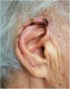
An 85-year-old woman from a remote region of Colombia (Orinoco region), presented with invasive basal cell carcinoma measuring 15 × 15 mm on the upper part of the right helix. The tumor was treated by transfixing resection with lateral margins of 8 mm. The postsurgical defect measured 35 mm in diameter and covered more than one-third of the length of the helix. Use of several flaps in different procedures was ruled out given the patient’s difficulty to travel to our hospital. Instead, the Antia-Buch technique was chosen using 2 flaps, an upper one and a lower one. To construct the flaps, an incision was made in the skin anterior to the ear and the cartilage without cutting posterior skin. Dissection was performed superficially to the posterior perichondrium, such that the flaps only had cartilage in their distal portion. A wedge was cut in the antihelix to enable the flaps to be drawn together. The flaps were advanced and sutured in layers with Vicryl® and Prolene® 5/0 stitches. A V-Y closure was performed on the upper secondary defect (Fig. 1). The outcome 1 week later was a functional pinna with no cup ear deformity (Fig. 2). The patient had an area of superficial necrosis of less than 1 cm that fully re-epithelialized.

Figure 1.

Surgical defect after resection (a). Positioning and suture of the flaps (b).

(0.11MB).
Figure 2.

One week after the operation.

(0.09MB).

Reconstruction of the pinna should seek to preserve its auxiliary function in the auditive process and as a support for glasses.2 The main esthetic objectives of reconstruction include retaining symmetry, preserving a smooth helix with its characteristic form, and preserving skin color and texture.4

There are different reconstructive techniques available and the choice of which to use depends on the site and size of the defects. In the upper helix, defects with no cartilage involvement can be resolved with skin grafts or local skin flaps. Defects of the helix with loss of cartilage may impact the shape of the pinna and so re-epithelization by second intent, grafts, and local skin flaps are insufficient.4 Helical defects of less than 10 mm can be resolved by primary closure or compound grafts.3 Larger defects need chondrocutaneous advancement flaps, such as the Antia-Buch flap, and, if the defects are greater than 20 mm, retroauricular island flaps or temporoparietal fascial flaps with cartilage graft can be used.3

Chondrocutaneous advancement flap of the helix is a simple technique described in 1967 by Antia and Buch to reconstruct the transfixing defects of the helix in a single surgical procedure.5 Depending on the size and site of the defect, 1 or 2 simultaneous flaps may be required. The incisions should be made parallel to the helix in the anterior face of the pinna, including the cartilage, but without making an incision in the posterior skin as the flaps are based on branches of the posterior auricular artery.6 The posterior face of the ear is then dissected to separate the skin from the cartilage.2 This maneuver frees the flaps and allows them to be advanced. Before suturing the flaps, it is necessary to assess whether they are long enough to surround the pinna. If not, cartilage should be cut away from the concha or the antihelix. The secondary defect of the upper flap can be closed with the V-Y technique,7 whereas the inferior flap may require a Burrow triangle to compensate for any redundant tissue (Fig. 3).2

Figure 3.

Step-by-step schematic of the Antia-Buch technique for the upper pole: lesion in the upper pole of the ear (a). Defect after tumor resection (b). Flap design (c). Dissection of the chondrocutaneous advancement flaps (d). Positioning and suture of the flaps (e).

(0.09MB).

The Antia-Buch technique plays an important role in reconstruction of the upper pole of the ear; however, more complex techniques are preferred for defects measuring more than 20 mm.3 One of the concerns with the Antia-Buch flap for large defects is cup ear deformity. However, modifications to this technique, such as half-moon or wedge cuts of the antihelix and concha can avoid this problem.2,7,8 A main advantage of this technique is that it only requires one operation. This is desirable in patients of advanced age, those with comorbidities,7 or those with difficulties traveling to hospital; 3 conditions often seen in our everyday practice. With this case, we would like to highlight the simplicity and usefulness of this technique for reconstruction of large defects of the pinna in the upper pole, particularly when only one operation is desirable.

Conflicts of interest

The authors declare that they have no conflicts of interest.

References
[1]
M.G. Pérez-Paredes, H.A. Cucunubo-Blanco, B. González-Sixto, M. Rodríguez Prieto.
Chondrocutaneous island flap for reconstruction of an antihelix defect.
Actas Dermosifiliogr, 106 (2015), pp. 503-504
[2]
A.L. Krunic, S. Weitzul, R.S. Taylor.
Chondrocutaneous advancement flap for reconstruction of helical rim defects in dermatologic surgery.
Australas J Dermatol, 47 (2006), pp. 296-299
[3]
M. Hénoux, F. Espitalier, A. Hamel, B. Dréno, G. Michel, O. Malard.
Vascular supply of the auricle: anatomical study and applications to external ear reconstruction.
Dermatol Surg, 43 (2017), pp. 87-97
[4]
D.G. Brodland.
Advanced reconstruction of the ear.
Dermatol Surg, 40 (2014), pp. S71-85
[5]
N.H. Antia, V.I. Buch.
Chondrocutaneous advancement flap for the marginal defect of the ear.
Plast Reconstr Surg, 39 (1967), pp. 472-477
[6]
H.J. De Schipper, J.H. van Rappard, E.A. Dumont.
Modified Antia Buch repair for full-thickness middle auricular defect.
Dermatol Surg, 38 (2012), pp. 124-127
[7]
A. Bialostocki, S.T. Tan.
Modified Antia-Buch repair for full-thickness upper pole auricular defects.
Plast Reconstr Surg, 103 (1999), pp. 1476-1479
[8]
E.M. Valesky, R. Kaufmann, M. Meissner.
Chondrocutaneous helix rim advancement flap: two classical and two new modifications for very large defects of the ear.
J Eur Acad Dermatol Venereol, 29 (2015), pp. 2248-2251

Please cite this article as: Barrera Gamboa JC, Acosta Madiedo de Hart AE. Colgajo de Antia-Buch para un defecto amplio del polo superior de la oreja. Actas Dermosifiliogr. 2021;112:558–559.

Copyright © 2020. AEDV
Descargar PDF
Idiomas
Actas Dermo-Sifiliográficas
Opciones de artículo
Herramientas
es en

¿Es usted profesional sanitario apto para prescribir o dispensar medicamentos?

Are you a health professional able to prescribe or dispense drugs?