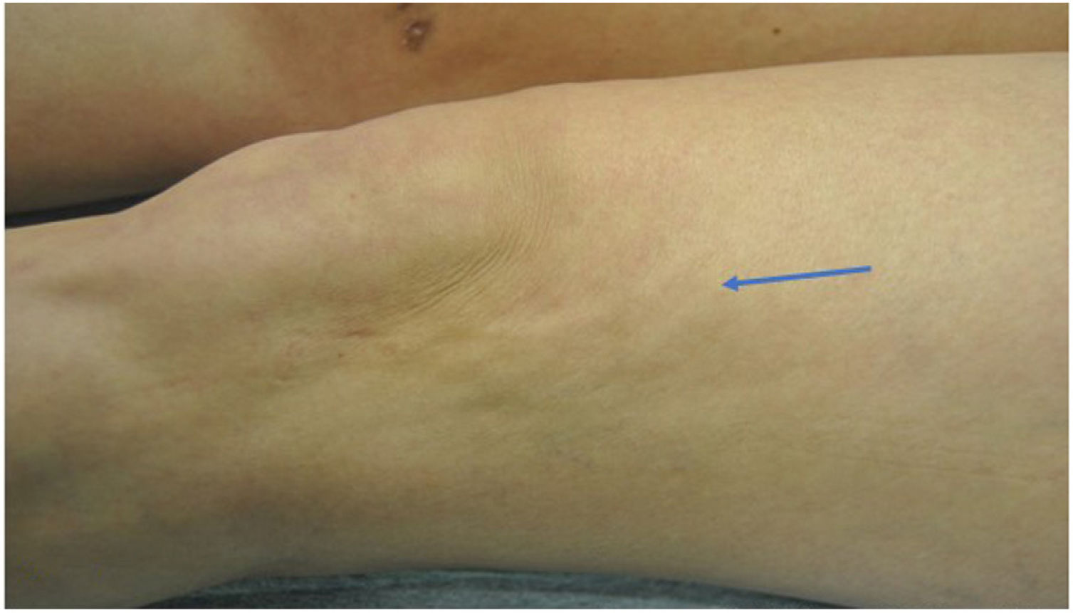
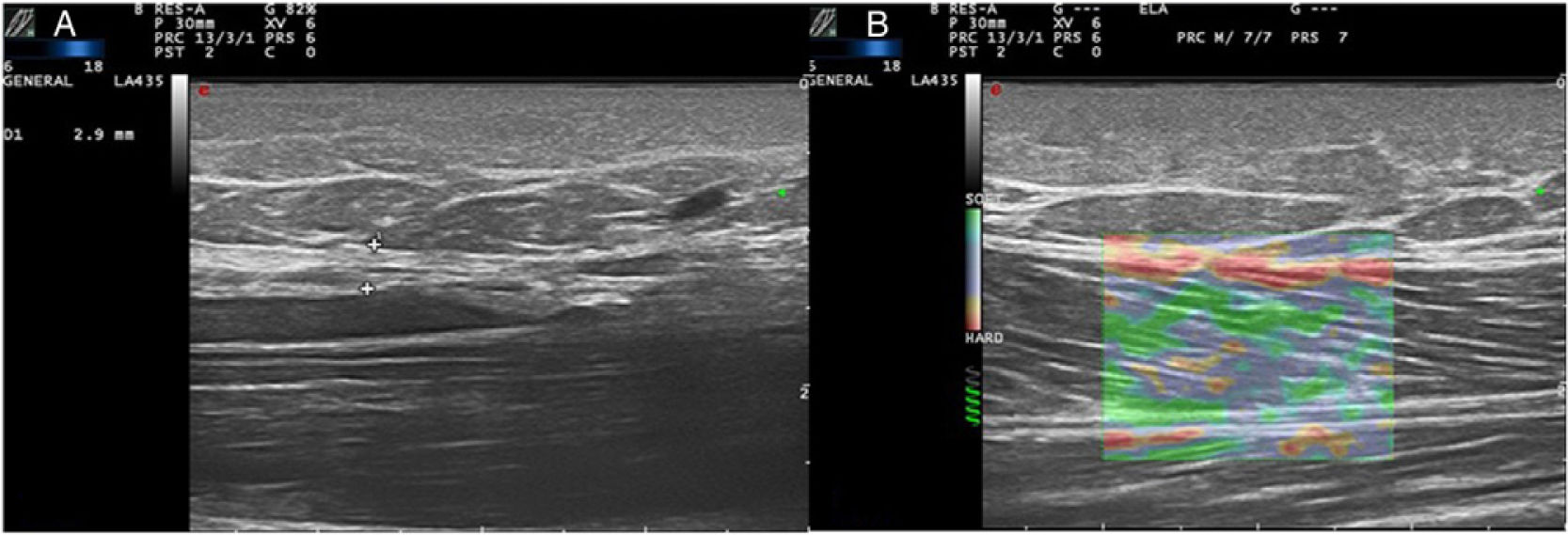
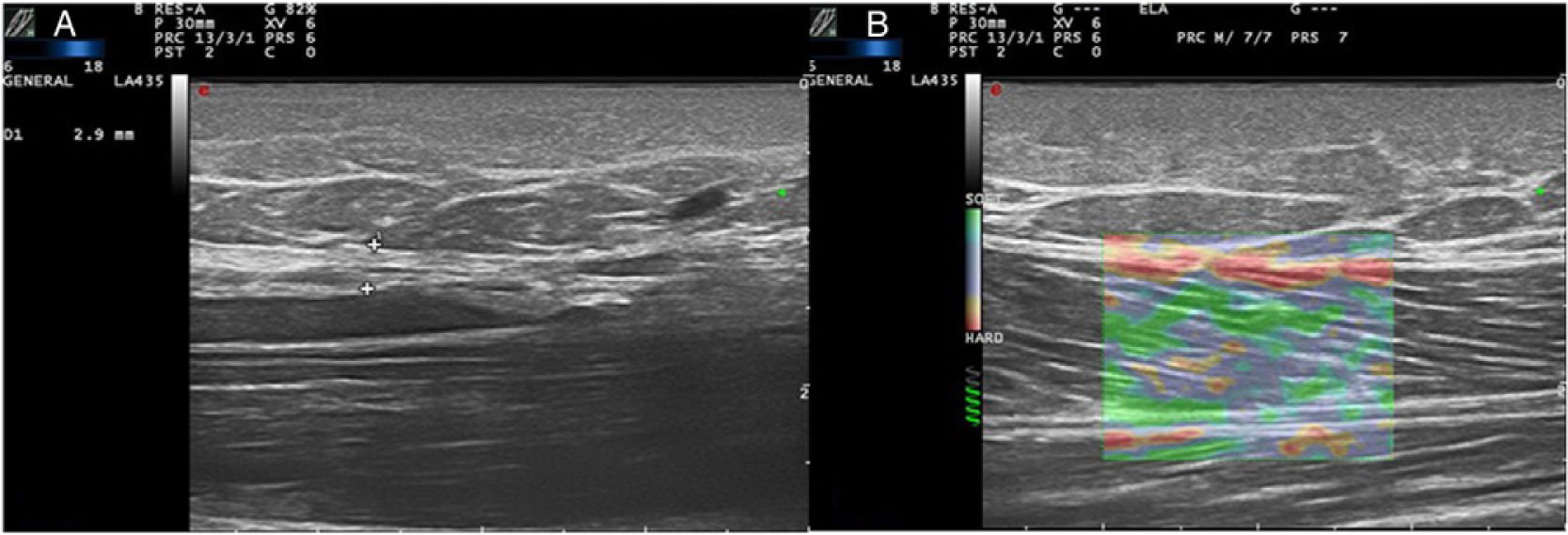
A 32-year-old woman presented with induration of the arms and the external aspect of the knees that had begun 3 months earlier and limited her movement (Fig. 1). Skin ultrasound (18-MHz linear probe, MyLab Class C, Esaote) revealed thickening of the muscular fascia, alterations of the echostructure of the superficial hypodermis, and a dermis with no pathological findings (Fig. 2A). Elastography showed increased stiffness of the muscle fascia (Fig. 2B). Based on these findings eosinophilic fasciitis (EF) was suspected, and was subsequently confirmed by nuclear magnetic resonance imaging and fascia biopsy. EF is a rare fibrosing disorder that manifests as pseudocellulitis (peau d’orange appearance), together with groove sign. Definitive diagnosis requires biopsy, which should include both the hypodermis and muscle fascia. Few articles have evaluated the diagnostic utility of ultrasound in EF. Ultrasound reveals an increase in the thickness of the muscular fascia, accompanied or not by alterations in the hypodermis. Elastography is a technique based on the detection of changes in tissue elasticity using ultrasound. Cutaneous ultrasound with elastography proved useful in guiding the diagnosis of this case of EF, and could potentially be used in follow-up to monitor treatment response.
Please cite this article as: Benavente Villegas FC, Alfageme Roldán F, Suárez Massa D. Signo de piel de naranja (seudocelulitis) y elastografía. Actas Dermosifiliogr. 2020;111:612.