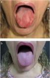
A 59-year-old woman consulted with a 2-year history of burning erythematous lesions on her tongue. Physical examination revealed glossitis, papillary atrophy, and erythematous macules on the dorsum and lateral borders (Fig. 1), hard palate, and oral mucosa. Histopathology revealed an inflammatory infiltrate with eosinophils and vascular congestion. Immunofluorescence was negative. Six months later, she presented again complaining of tiredness, paresthesia in the lower limbs, and persistence of the oral lesions.
The laboratory work-up revealed macrocytic anemia with a normal reticulocyte count, pancytopenia, and increased lactate dehydrogenase and indirect bilirubin. The findings of positive intrinsic factor antibody titers confirmed the diagnosis of pernicious anemia.
The patient was prescribed intramuscular hydroxocobalamin (1000μg/d for 7 days, followed by 1000μg/wk for 4 weeks). The oral lesions resolved after 1 month of treatment (Fig. 1B), and her complete blood count was within normal values. The patient continued to receive monthly injections of hydroxocobalamin.
Pernicious anemia is the deficiency of cobalamin caused by intrinsic factor antibodies, which interfere with its absorption and lead to ineffective erythropoiesis. Oral abnormalities such as glossitis, papillary atrophy, erythematous macules, angular cheilitis, and burning tongue pain precede the hematological abnormalities. The present case highlights the importance of a laboratory work-up for identification of this disease in patients with burning oral pain and skin lesions.
Please cite this article as: Gonzalez-Benavides N, Rodriguez-Vivian C, Ocampo-Candiani J. Máculas eritematosas urentes orales como manifestación inicial de deficiencia de la vitamina B12. Actas Dermosifiliogr. 2020;111:331–331.


