Journal Information
Vol. 105. Issue 3.
Pages 243-252 (April 2014)
Visits
16393
Vol. 105. Issue 3.
Pages 243-252 (April 2014)
Review
Full text access
Antimalarials in Dermatology: Mechanism of Action, Indications, and Side Effects
Antipalúdicos en dermatología: mecanismo de acción, indicaciones y efectos secundarios
Visits
16393
C. Rodriguez-Caruncho
Corresponding author
clararodriguezc@yahoo.es

Corresponding author.
, I. Bielsa Marsol
Servicio de Dermatología, Hospital Universitari Germans Trias i Pujol, Universitat Autónoma de Badalona, Badalona, Barcelona, Spain
This item has received
Article information
Abstract
Full Text
Bibliography
Download PDF
Statistics
Tables (5)
Table 1. Mechanisms of Action of Antimalarial Agents.
Table 2. Indications for Antimalarial Agents: Miscellaneous Conditions.
Table 3. Risk Factors for Retinopathy with Antimalarial Therapy.
Table 4. Drug-Drug Interactions Involving Antimalarial Agents.
Table 5. Contraindications of Antimalarial Agents.
Show moreShow less
Abstract

Antimalarial drugs have been in common use in dermatology since the 1950s. Their mechanism of action is complex, and it is now known that they act through various pathways. We review the indications for antimalarials in dermatology, their adverse effects, and some less well-known effects, such as their antithrombotic and hypolipidemic action. The most recent recommendations concerning ophthalmological screening in patients on antimalarials are also reviewed.

Keywords:
Antimalarials
Chloroquine
Hydroxychloroquine
Lupus erythematosus
Resumen

Los antipalúdicos (AP) son fármacos de uso habitual en dermatología desde la década de los 50. Su mecanismo de acción es complejo, y actualmente se sabe que actúan por diversas vías. En este artículo se revisan las indicaciones de los antimaláricos en dermatología, sus efectos secundarios y algunos efectos menos conocidos, como el antitrombótico o el hipolipidemiante. Se recogen también las recomendaciones más recientes acerca del seguimiento oftalmológico de estos pacientes.

Palabras clave:
Antipalúdicos
Cloroquina
Hidroxicloroquina
Lupus eritematoso
Full Text

Antimalarial drugs have been known for more than 300 years. The antipyretic properties of bark from cinchona, a tree native to South America, were already known in the 17th century. The first natural antimalarial agent, quinine, was obtained from this tree and was used by European colonists in tropical countries as protection against malaria. The beneficial effects of quinine in patients with lupus erythematosus (LE) were published in 1894 by Payne, who reported the successful treatment of discoid LE with this drug.1 The first synthetic antimalarial, quinacrine (QC), was made in 1930. Chloroquine (CQ) and hydroxychloroquine (HCQ) followed (CQ was first synthesized in 1934 and HCQ in 1955).2 The use of antimalarials as a treatment for LE became widespread in 1951, with the publication of an article by Page,3 which described good response to QC treatment in 18 patients with LE.

The antimalarials used in dermatology are CQ, HCQ, and QC. The latter of these is not commercially available in Spain and can only be used by making an application for a foreign medicine.

Pharmacokinetics

CQ and HCQ are 4-aminoquinoleins which differ from each other in that HCQ has a hydroxylated side chain. CQ is formulated as a diphosphate for oral administration. Between 90% and 100% is absorbed in the gastrointestinal tract. Stable plasma concentrations are reached after 4 to 6 weeks, and so most cases require drug administration for at least this duration to obtain therapeutic response. CQ binds strongly to plasma proteins which are deposited in tissues such as the liver, spleen, kidneys, and lungs, and in blood cells.4,5 Avidity is particularly high for skin and melanin-containing retinal cells, where concentrations are between 100 and 200 times the plasma concentration. Between 45% and 50% of these plasma proteins are eliminated in urine. CQ has a long half-life, which can vary between 74 hours and 50 days depending on the cumulative dose. The drug can remain in the skin for 6 to 7 months after discontinuation of therapy.6 Both drugs cross the placenta and are excreted in small amounts in breast milk.7

QC is a 9-aminocridin and its pharmacokinetics are similar to those of the 4-aminoquinoleins. It is also quickly absorbed after oral administration and steady-state concentrations are reached after 4 weeks.

Mechanism of Action

The mechanism of action has not been fully elucidated, but they are known to act on a range of pathways and have immunomodulatory, anti-inflammatory, antiproliferative, and photoprotective effects. Antimalarials are weak bases that can readily cross cell membranes and accumulate in acidic cytoplasmic vesicles (lysosomes or endosomes), where they remain trapped in a protonated state.8 This mechanism of action places antimalarials in the therapeutic group known as lysosomotropic drugs.9 The pH in lysosomes increases as antimalarials accumulate there, subsequently interfering in binding of antigenic peptides with class II molecules of the major histocompatibility complex. Presentation to CD4+ T lymphocytes is thus avoided, leading to inhibition of the production of cytokines that participate in the generation of inflammatory response.10,11 A mechanism has been proposed whereby antimalarials can act as immunomodulators without causing immunosuppression. According to this proposed mechanism, inhibition of binding with the major histocompatibility complex only occurs for autoantigens and not for exogenous peptides. Autoantigens are low affinity peptides. Thus, they do not bind to the alpha and beta chains of the major histocompatibility complex molecules when the pH of the vesicle increases. Exogenous peptides, in contrast, have a high affinity, and so binding does occur and they are presented to the T lymphocytes.12,13

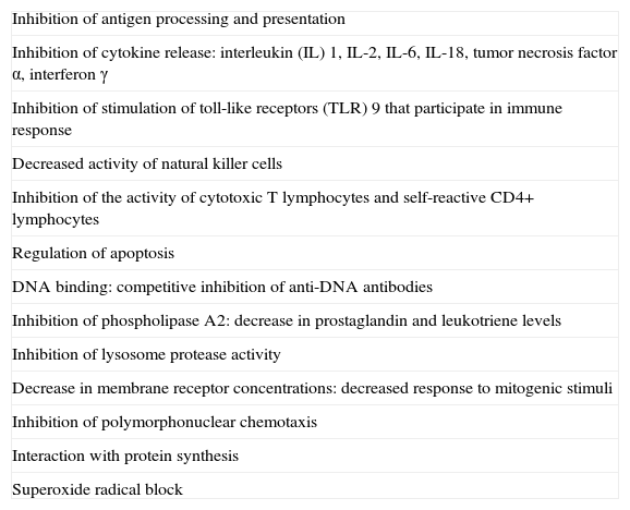
In addition, antimalarials act through other immunomodulatory, anti-inflammatory, and antiproliferative mechanisms (Table 1).10,14–18

Table 1.

Mechanisms of Action of Antimalarial Agents.

Inhibition of antigen processing and presentation 
Inhibition of cytokine release: interleukin (IL) 1, IL-2, IL-6, IL-18, tumor necrosis factor α, interferon γ 
Inhibition of stimulation of toll-like receptors (TLR) 9 that participate in immune response 
Decreased activity of natural killer cells 
Inhibition of the activity of cytotoxic T lymphocytes and self-reactive CD4+ lymphocytes 
Regulation of apoptosis 
DNA binding: competitive inhibition of anti-DNA antibodies 
Inhibition of phospholipase A2: decrease in prostaglandin and leukotriene levels 
Inhibition of lysosome protease activity 
Decrease in membrane receptor concentrations: decreased response to mitogenic stimuli 
Inhibition of polymorphonuclear chemotaxis 
Interaction with protein synthesis 
Superoxide radical block 

Finally, a photoprotective effect has been attributed to these drugs, although the mechanism is not yet understood. One possibility is that these drugs may have a certain screen effect, absorbing certain wavelengths of sunlight.17 Another is that antimalarials inhibit the inflammatory response of keratinocytes which is triggered by exposure to sunlight through induction of apoptosis and subsequent exposure to keratinocyte antigens.11 Moreover, the drug may enhance natural photoprotection of the epidermis through induction of c-jun transcription.19

Other Antimalarial Actions

Other actions have also been attributed to antimalarials. These include antithrombotic, lipid-lowering, and glucose-lowering effects and effects on bone metabolism. The studies cited below have been conducted mainly in patients with systemic lupus erythematosus (SLE), and in most cases the agent used was HCQ.

Antithrombotic Action

The antithrombotic effect of antimalarials has been attributed to a range of mechanisms. First, a reduction in red blood cell aggregation has been observed.20 Second, antimalarials inhibit platelet aggregation,21,22 and might also reduce blood viscosity.23 Antimalarials also hinder platelet aggregation induced by antiphospholipid antibody,24 and inhibit the production of thromboxane A2 through deactivation of phospholipase A2 and prostaglandins in platelet membranes.25 In addition, some authors have suggested that these drugs have a synergistic antiatherosclerotic effect which prevents thrombus formation, although the strength of scientific evidence in this case is limited.26

The antithrombotic action of antimalarials has been known since the 1970s, when some antimalarials were investigated for the prevention of thrombosis after surgery.27–29 In these studies, however, the doses of HCQ used ranged from 600 to 1000mg, which is considerably higher than those usually used in patients with LE.

Since the end of the 1980s, numerous studies have been performed to investigate the possible antithrombotic effect of antimalarials in patients with SLE both with and without associated antiphospholipid syndrome.30–38 In general, these studies indicate that antimalarials have an effect on the prevention of thrombotic phenomena in patients with SLE. There are insufficient data to draw conclusions about whether the antithrombotic effect is greater for arterial or venous events.26

Lipid-Lowering Effect

Several studies have been performed to assess the effect of antimalarials on the lipid profile.39–47 In all except 2,43,47 there were significant differences between patients who were taking antimalarials and those who were not. Effects on levels of total cholesterol, low-density lipoprotein cholesterol, very-low-density lipoprotein cholesterol, and triglycerides have been observed.41,42

Glucose-Lowering Effect

Patients with SLE treated with antimalarials have shown a trend towards better control of glucose levels, decreased insulin resistance, and lower levels of hemoglobin A1c.48–50

Effects on Bone Metabolism

Two studies with small cohorts of patients with SLE have investigated the effect of HCQ on bone mineral density.51,52 In both studies, the patients treated with HCQ had a greater bone mineral density measured in the spine51 and in the hips.52

Dosing

For most indications, including LE, the normal maintenance doses of CQ and HCQ are 250mg/d and 200mg/d, respectively. These doses are usually doubled in the first 15 to 30 days of drug administration and in the event of lesion flare-ups. QC, which is not usually used in Spain, is administered at a dose of 100mg/d.

Formulas are available to adjust the daily dose that can be administered to reduce the risk of retinopathy. These doses are calculated according to the ideal weight of the patient. Examples of these formulas for calculating this ideal weight are as follows: for women (height in cm - 100) - 15% and for men (height in cm - 100) - 10%. If the patient weighs less than the ideal weight, the doses are adjusted according to the real weight. The dose to be administered is calculated as follows: 3.5-4mg × ideal body weight in kg for CQ and 6-6.5mg × ideal body weight in kg for HCQ. With these formulas, the HCQ dose will easily surpass 200mg/d; in this case, 400mg/d can be administered on the number of days of the week necessary. For example, for an ideal weight of 40-43kg, 400mg/d is administered twice a week and 200mg/d for the remaining days.53 In the case of CQ, in patients with a low ideal body weight, the daily dose will be below 250mg/d. Given that tablets with a dose less than 250mg are not available in Spain, one possibility would be not to take the drug one or several days of the week.

Dose calculation using these formulas is particularly important for long-term treatment and in small patients, in whom there is a risk of overdose with the standard antimalarial doses. These doses should be reduced and individualized in patients with impaired liver or renal function.53

Indications in Dermatology

For practical purposes, we can divide the indications of antimalarials into indications as first-line treatment, indications as second- or third-line treatments, and miscellaneous (use in isolated clinical cases). The indications of antimalarials in rheumatologic diseases such as rheumatoid arthritis or Sjörgren syndrome will not be discussed here.

Indications As First-Line DrugsLupus Erythematosus

LE is the only dermatologic indication approved by the US Food and Drug Administration.

Antimalarials are effective for the treatment of the specific skin lesions of cutaneous LE (CLE), whether acute, subacute, or chronic.54-56 However, in the case of chronic CLE, a worse response has been observed in the hypertrophic and verrucous forms, disseminated lesions, and lesions associated with SLE.10,56 Lesions do respond well in cases of tumid LE, even with the chronic forms.57 In patients with SLE, antimalarials also improve certain systemic manifestations, such as arthralgia, myalgia, and serositis.58 They also have an effect on nonspecific lesions of the disease, such as oral ulcers, photosensitivity, and calcinosis cutis.59 In patients with CLE, it is normal clinical practice for patients to follow intermittent treatments, with antimalarials administered during the months of highest exposure to sunlight, when flare-ups are most frequent, and suspended during fall and winter. The drug is therefore suspended in those periods of inactivity of the cutaneous disease.

In cases of LE in which there is no response to the first-line antimalarial treatment, a switch to another antimalarial can be tried (from HCQ to CQ or vice versa). In patients with refractory cutaneous disease, a combination of antimalarials may be appropriate, combining QC with any of the other 2 antimalarials.60 The action of QC is not associated with a greater risk of retinal toxicity.

Smoking has been shown to be associated with decreased efficacy of antimalarials in several studies,61–63 although in a recent retrospective study the authors found no relationship between smoking and HCQ response.56

Porphyria Cutanea Tarda

Phlebotomy is the mainstay treatment for porphyria cutanea tarda. Nevertheless, in certain cases, such as in patients who do not response or in whom this procedure is contraindicated, antimalarials can be administered.64–66 Antimalarials enhance hepatic depletion and urinary excretion of porphyrins while also inhibiting their synthesis.18 The doses used should be low to avoid massive porphyrin depletion and liver damage. Therefore, a dose of 2mg/kg twice a week is recommended in the case of CQ and of 3.5mg/kg twice a week in the case of HCQ.10,17,18 This dose is usually slowly increased according to response. Some authors recommend administering a test dose of 125-250mg of CQ and performing an analysis of liver enzymes before continuing with treatment.67

According to some studies, 1 to 4 phlebotomies, performed prior to administration of the antimalarials, may enhance drug response and reduce the risk of liver damage.68,69

Chronic Ulcerative Stomatitis

Chronic ulcerative stomatitis (CUS) was first described as an entity in 1990.70,71 The disease occurs mainly in elderly women and follows a course of painful ulceration in the oral mucosa. The clinical and pathologic features are similar to erosive lichen planus. The disease has been shown to be caused by antinuclear antibodies reactive exclusively to the squamous epithelium and specifically to the delta Np64 alpha protein, expressed in epithelial basal cells.72–75

A characteristic feature of CUS is that it is refractory to topical and systemic corticosteroids. In contrast, CUS responds well to antimalarials, and so these are the first-line treatment.18

Indications as Second- and Third-line TreatmentsDermatomyositis

Antimalarials have been shown to be effective in the treatment of dermatomyositis with substantial skin involvement. They are administered either as monotherapy in cases of dermatomyositis without muscle weakening or as a coadjuvant treatment in those with associated muscle disease.76–80 Although antimalarials mainly improve the skin manifestations, they have been shown to also improve muscle strength in combination with oral corticosteroids.81 In patients who do not respond to monotherapy with antimalarials, combined therapy with QC can be administered.79 It should be pointed out that patients with dermatomyositis have a higher risk of cutaneous side effects from taking antimalarials.82

Sarcoidosis

Systemic or topical corticosteroids are the first-line treatment for sarcoidosis. However, antimalarials are effective in the treatment not only of skin manifestations of this disease83–86 but also of pulmonary manifestations87–89 and certain systemic complications.90–94

Polymorphous Light Eruption

Although antimalarials are not the first-line treatment for polymorphous light eruption (PLE), several studies have reported good therapeutic outcomes with these drugs.95–97 Two controlled studies have found an increased tolerance to sunlight exposure and a decrease in rash; itching, however, was alleviated to a lesser extent.98,99

Antimalarials are indicated in patients with PLE who do not respond to usual therapy, such as photoprotection, hardening associated with phototherapy, and topical corticosteroids.10,18 Some authors suggest starting HCQ at a dose of 400mg/d from a few days prior to exposure to sunlight and reducing the dose to 200mg/d once clinical stability is attained.18

Disseminated Granuloma Annulare

Publications on the use of antimalarials in disseminated granuloma annulare are limited to a few case series.100–102 However, use of antimalarials is recommended in patients with disseminated granuloma annulare that does not respond to topical corticosteroids or when corticosteroids cannot be used due to the extension of the lesions.10,18

Miscellaneous Conditions

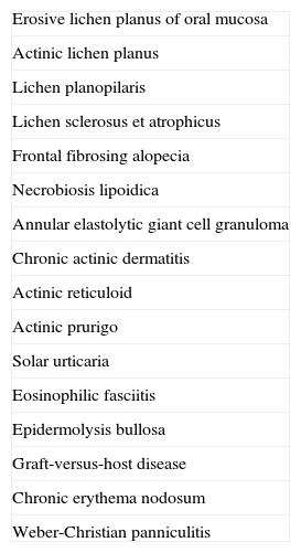
Table 2 summarizes some conditions in which antimalarials have been used, although the scientific evidence is scant and limited to the publication of isolated case reports.103–119

Table 2.

Indications for Antimalarial Agents: Miscellaneous Conditions.

Erosive lichen planus of oral mucosa 
Actinic lichen planus 
Lichen planopilaris 
Lichen sclerosus et atrophicus 
Frontal fibrosing alopecia 
Necrobiosis lipoidica 
Annular elastolytic giant cell granuloma 
Chronic actinic dermatitis 
Actinic reticuloid 
Actinic prurigo 
Solar urticaria 
Eosinophilic fasciitis 
Epidermolysis bullosa 
Graft-versus-host disease 
Chronic erythema nodosum 
Weber-Christian panniculitis 
Side Effects, Contraindications, and InteractionsSide EffectsOcular Effects

Antimalarials can cause several reversible ocular side effects. Retinopathy is the most feared side effect of these agents and can, at times, lead to an irreversible vision loss.

Reversible ocular effects include corneal deposits, which can be asymptomatic or cause blurred vision and halos of colors around lights. These deposits are observed in up to 90% of patients who take CQ and 5% of those who take HCQ.120 There is no direct relationship with retinopathy, but the presence of such deposits can be a warning for the need for closer follow-up. Accomodation disorders and diplopia are other reversible ocular side effects of these agents. Diplopia occurs more frequently with CQ and usually resolves in time or with a decrease in dose.

Retinopathy is the most serious ocular side effect and, if detected, treatment should be suspended. Antimalarials are known to have avidity for melanin in the pigmentary epithelium of the retina; however, the mechanism by which retinopathy occurs is not well known.121 In the early stages, functional loss in the paracentral region of the retina may occur. The screening methods used should be able to detect patients in this stage, at which vision loss does not progress if the drug is withdrawn. If exposure to the drug continues, the subsequent retinopathy gives rise to an image with a bull's eye appearance, which consists of macular paleness surrounded by several rings. In this case, the damage is irreversible. Depigmentation often occurs along with progressive functional loss up to 1 year after suspending treatment. Advanced cases show diffuse retinal atrophy associated with loss of visual acuity, peripheral vision, and night vision.121

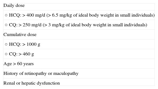
The prevalence of retinopathy caused by antimalarials is low, particularly in the first 5 years of treatment. However, with longer treatment durations, it can rise to 1%.122 The risk of ocular toxicity thus increases in patients with cumulative doses greater than 1000 g of HCQ and 460 g of CQ. This risk is also greater in patients who exceed the dose of 3.5-4mg/kg of ideal body weight for CQ and 6-6.5mg/kg of × ideal body weight for HCQ.121 As mentioned above, these daily doses are not exceeded in most patients who take 400mg/d of HCQ or 250mg/d of CQ, but small patients may exceed this threshold. However, we should bear in mind that cases of retinal toxicity have been described despite taking doses below these limits and for a short period of time.123 In general, it is accepted that the risk of developing retinopathy is greater in patients treated with CQ than in those treated with HCQ, while the risk in those receiving QC is almost nonexistent.26,124 However, the recommendations for screening of the American Academy of Ophthalmology (AAO) are the same for CQ and HQC. The most recent update of these recommendations, published in 2011, recommend baseline ocular control using biomicroscopy, automated perimetry, fundus examination and at least one of the following tests: spectral domain optical coherence tomography, fundus autofluorescence imaging, and multifocal electroretinogram. Annual screening should start after 5 years of treatment with antimalarials in patients without any risk factors, and from the start of treatment in those patients with the risk factors shown in Table 3. According to the AAO, annual screening should include automated perimetry and, if possible, at least 1 of the 3 aforementioned tests: spectral domain optical coherence tomography, fundus autofluorescence imaging, and multifocal electroretinogram.121 The authors of this review insist, however, that these are the minimum number of recommended controls and the treating physician is free to increase the frequency of these visits or the number of tests ordered.

Table 3.

Risk Factors for Retinopathy with Antimalarial Therapy.

Daily dose 
○ HCQ:>400mg/d (>6.5mg/kg of ideal body weight in small individuals) 
○ CQ:>250mg/d (>3mg/kg of ideal body weight in small individuals) 
Cumulative dose 
○ HCQ:>1000 g 
○ CQ:>460 g 
Age>60 years 
History of retinopathy or maculopathy 
Renal or hepatic dysfunction 
Gastrointestinal Effects

Gastrointestinal effects are the most common reactions to antimalarials, although they are usually mild in severity and can be managed by dose reduction. They occur most frequently in patients treated with QC (30%), followed by those who take CQ (20%) and HCQ (10%).2 The most common gastrointestinal events are nausea, vomiting, and diarrhea. Less frequent gastrointestinal events include anorexia, abdominal distension, and transaminitis.10

Cutaneous Effects

Some patients who take antimalarials in the long term develop a grey-blue skin pigmentation, which is more evident on the face, palate, forearms, and legs. QC causes darker pigmentation than CQ and HCQ. In addition, the hair roots may become whitened and transversal bands may appear in the nails.125 These disorders resolve a few months after suspending treatment.

Other cutaneous side effects are pruritus, rash, erythroderma, exfoliative dermatitis, urticaria, eczema, alopecia, photosensitivity, and erythema annulare centrifugum.10

Neuromuscular Effects

Antimalarials have been reported to cause some psychiatric side effects, such as psychosis, irritability, depression, insomnia, and nightmares. These events, however, usually occur in patients treated at higher doses than those used in dermatology.126,127 Antimalarials can also induce seizure in predisposed individuals,128 and cause myotoxicity due to the accumulation of the drug in the vacuoles of the myofibrils of the striated muscle. This myopathy, which affects the proximal muscles and may be accompanied by peripheral neuropathy, usually resolves with discontinuation of the antimalarial.129

Cardiac Effects

Occasionally, antimalarials can cause conduction disorders and hypertrophic cardiomyopathy.10,130 However, 2 studies that specifically assessed cardiotoxicity in patients treated with antimalarials did not find any significant cardiac side effects.131,132

Hematologic Effects

Hematologic side effects are uncommon. Hemolysis in patients with glucose-6-phosphate dehydrogenase deficiency, aplastic anemia, and leukopenia has been reported.10 These disorders occur more frequently in patients treated with QC, especially when used at higher doses.133 The need for blood tests during treatment is subject to debate, although testing is generally not considered necessary unless the patient has an underlying hematologic disease.

Drug-Drug Interactions

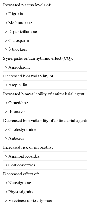
Antimalarials can interact with some drugs. These interactions are listed in Table 4.

Table 4.

Drug-Drug Interactions Involving Antimalarial Agents.

Increased plasma levels of: 
○ Digoxin 
○ Methotrexate 
○ D-penicillamine 
○ Ciclosporin 
○ β-blockers 
Synergistic antiarrhythmic effect (CQ): 
○ Amiodarone 
Decreased bioavailability of: 
○ Ampicillin 
Increased bioavailability of antimalarial agent: 
○ Cimetidine 
○ Ritonavir 
Decreased bioavailability of antimalarial agent: 
○ Cholestyramine 
○ Antacids 
Increased risk of myopathy: 
○ Aminoglycosides 
○ Corticosteroids 
Decreased effect of: 
○ Neostigmine 
○ Physostigmine 
○ Vaccines: rabies, typhus 

Source: Kalia S et al.,10 Ochsendorf FR,53 Wozniacka A et al.133

Contraindications

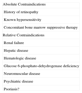
The absolute and relative contraindications of antimalarials are summarized in Table 5.

Table 5.

Contraindications of Antimalarial Agents.

Absolute Contraindications 
History of retinopathy 
Known hypersensitivity 
Concomitant bone marrow suppressive therapy 
Relative Contraindications 
Renal failure 
Hepatic disease 
Hematologic disease 
Glucose 6-phosphato-dehydrogenase deficiency 
Neuromuscular disease 
Psychiatric disease 
Psoriasis? 

Source: Bielsa I,17 Wozniacka A et al.133

Antimalarials and Pregnancy

Antimalarials can cross the placenta and reach the fetus.134 However, several studies have assessed the safety of these drugs during pregnancy and none have detected a greater risk of congenital malformations or ocular, neurologic, or auditory toxicity.135–141 The level of evidence for this absence of effect is greater for HCQ than for CQ, as HCQ was the drug administered to most of the pregnant women studied.26 In studies conducted in patients with SLE, the benefit of continuing treatment with antimalarials during pregnancy is generally acceptable because the risk of disease complications during pregnancy is greater than the risk the drug poses to the fetus.10

Ethical ResponsibilitiesProtection of human and animal subjects.

The authors declare that no experiments were performed on humans or animals for this investigation.

Confidentiality of data.

The authors declare that they have followed their hospital's protocol on the publication of data concerning patients and that all patients included in the study have received sufficient information and have given their written informed consent to participate in the study.

Right to privacy and informed consent.

The authors declare that patient data do not appear in this article.

Conflicts of Interest

Dr. Clara Rodriguez-Caruncho has received research grants from Laboratorios Rubió. I Bilsa Marsol declares that she has no conflicts of interest.

References
[1]
D.J. Wallace.
The history of antimalarials.
Lupus, 5 (1996), pp. S2-S3
[2]
M.J. Van Beek, W.W. Piette.
Antimalarials.
Dermatol Clin, 19 (2001), pp. 147-160
[3]
F. Page.
Treatment of lupus erythematosus with mepacrine.
Lancet, 2 (1951), pp. 755-758
[4]
J. Ducharme, R. Farinotti.
Clinical pharmacokinetics and metabolism of chloroquine. Focus on recent advancements.
Clin Pharmacokinet, 3 (1996), pp. 257-274
[5]
S. Krishna, N.J. White.
Pharmacokinetics of quinine, chloroquine and amodiaquine. Clinical implications.
Clin Pharmacokinet, 30 (1996), pp. 263-299
[6]
G. Sjölin-Forsberg, B. Berne, C. Blixt, M. Johansson, B. Lindström.
Chloroquine phosphate: a long-term follow-up of drug concentrations in skin suction blister fluid and plasma.
Acta Derm Venereol, 73 (1993), pp. 426-429
[7]
N. Costedoat-Chalumeau, Z. Amoura, P. Duhaut, D.L. Huong, D. Sebbough, B. Wechsler, et al.
Safety of hydroxychloroquine in pregnant patients with connective tissue diseases: a study of one hundred thirty-three cases compared with a control group.
Arthritis Rheum, 48 (2003), pp. 3207-3211
[8]
A.M. Kaufmann, J.P. Krise.
Lysosomal sequestration of amine-containing drugs: analysis and therapeutic implications.
J Pharm Sci, 96 (2007), pp. 729-746
[9]
C. De Duve, T. de Barsy, B. Poole, A. Trouet, P. Tulkens, F. van Hoof.
Commentary. Lysosomotropic agents.
Biochem Pharmacol, 23 (1974), pp. 495-531
[10]
S. Kalia, J.P. Dutz.
New concepts in antimalarial use and mode of action in dermatology.
Dermatol Ther, 20 (2007), pp. 160-174
[11]
A. Wozniacka, A. Carter, D.P. McCauliffe.
Antimalarials in cutaneous lupus erythematosus: mechanisms of therapeutic benefit.
Lupus, 11 (2002), pp. 71-81
[12]
R.I. Fox.
Mechanism of action of hydroxychloroquine as an antirheumatic drug.
Semin Arthritis Rheum, 23 (1993), pp. 82-91
[13]
R. Fox.
Anti-malarial drugs: possible mechanisms of action in autoimmune disease and prospects for drug development.
Lupus, 5 (1996), pp. S4-S10
[14]
M. Jiménez.
Antipalúdicos: actualización de su uso en enfermedades reumáticas.
Reumatol Clin, 2 (2006), pp. 190-201
[15]
R.I. Rynes.
Antimalarial drugs in the treatment of rheumatological diseases.
Br J Rheumatol, 36 (1997), pp. 799-805
[16]
J. Bondeson, R. Sundler.
Antimalarial drugs inhibit phospholipase A2 activation and induction of interleukin 1beta and tumor necrosis factor alpha in macrophages: implications for their mode of action in rheumatoid arthritis.
Gen Pharmacol, 30 (1998), pp. 357-366
[17]
I. Bielsa.
Uso de los antimaláricos en dermatología.
Piel, 18 (2003), pp. 515-518
[18]
R. Wolf, D. Wolf, V. Ruocco.
Antimalarials: unapproved uses or indications.
Clin Dermatol, 18 (2000), pp. 17-35
[19]
T.Q. Nguyen, J.D. Capra, R.D. Sontheimer.
4-Aminoquinoline antimalarials enhance UV-B induced c-jun transcriptional activation.
Lupus, 7 (1998), pp. 148-153
[20]
B.P. Madow.
Use of antimalarial drugs as desludging agents in vascular disease processes.
J Am Med Assoc, 172 (1960), pp. 1630-1633
[21]
E. Bertrand, B. Cloitre, R. Ticolat, R.K. Bile, C. Gautier, G.O. Abiyou, et al.
Antiaggregation action of chloroquine.
Med Trop (Mars), 50 (1990), pp. 143-146
[22]
P.D. Winocour, R.L. Kinlough-Rathbone, J.F. Mustard.
The effect of phospholipase inhibitor mepacrine on platelet aggregation, the platelet release reaction and fibrinogen binding to the platelet surface.
Thromb Haemost, 45 (1981), pp. 257-262
[23]
E. Ernst, M. Rose, R. Lee.
Modification of transoperative changes in blood fluidity by hydroxychloroquine: a possible explanation for the drug's antithrombotic effect.
Pharmatherapeutica, 4 (1984), pp. 48-52
[24]
R.G. Espinola, S.S. Pierangeli, A.E. Gharavi, E.N. Harris.
Hydroxychloroquine reverses platelet activation induced by human IgG antiphospholipid antibodies.
Thromb Haemost, 87 (2002), pp. 518-522
[25]
R. Nosál, V. Jancinová, M. Petríková.
Chloroquine inhibits stimulated platelets at the arachidonic acid pathway.
Thromb Res, 77 (1995), pp. 531-542
[26]
G. Ruiz-Irastorza, M. Ramos-Casals, P. Brito-Zeron, M.A. Khamashta.
Clinical efficacy and side effects of antimalarials in systemic lupus erythematosus: a systematic review.
Ann Rheum Dis, 69 (2010), pp. 20-28
[27]
D.B. Pilcher.
Hydroxychloroquine sulfate in prevention of thromboembolic phenomena in surgical patients.
Am Surg, 41 (1975), pp. 761-766
[28]
J.R. Loudon.
Hydroxychloroquine and postoperative thromboembolism after total hip replacement.
Am J Med, 85 (1988), pp. 57-61
[29]
E. Johansson, K. Forsberg, H. Johnsson.
Clinical and experimental evaluation of the thromboprophylactic effect of hydroxychloroquine sulfate after total hip replacement.
Haemostasis, 10 (1981), pp. 89-96
[30]
D.J. Wallace.
Does hydroxychloroquine sulfate prevent clot formation in systemic lupus erythematosus.
Arthritis Rheum, 30 (1987), pp. 1435-1436
[31]
D. Erkan, Y. Yazici, M.G. Peterson, L. Sammaritano, M.D. Lockshin.
A cross-sectional study of clinical thrombotic risk factors and preventive treatments in antiphospholipid syndrome.
Rheumatology (Oxford), 41 (2002), pp. 924-929
[32]
S.M. Toloza, A.G. Uribe, G. JJr. McGwin, G.S. Alarcón, B.J. Fessler, H.M. Bastian, et al.
Systemic lupus erythematosus in a multiethnic US cohort (LUMINA). XXIII. Baseline predictors of vascular events.
Arthritis Rheum, 50 (2004), pp. 3947-3957
[33]
C.C. Mok, S.S. Tang, C.H. To, M. Petri.
Incidence and risk factors of thromboembolism in systemic lupus erythematosus: a comparison of three ethnic groups.
Arthritis Rheum, 52 (2005), pp. 2774-2782
[34]
K.T. Ho, C.W. Ahn, G.S. Alarcón, B.A. Baethge, F.K. Tan, J. Roseman, et al.
Systemic lupus erythematosus in a multiethnic cohort (LUMINA): XXVIII. Factors predictive of thrombotic events.
Rheumatology (Oxford), 44 (2005), pp. 1303-1307
[35]
K. De Leeuw, B. Freire, A.J. Smit, H. Bootsma, C.G. Kallenberg, M. Bijl.
Traditional and non-traditional risk factors contribute to the development of accelerated atherosclerosis in patients with systemic lupus erythematosus.
Lupus, 15 (2006), pp. 675-682
[36]
G. Ruiz-Irastorza, M.V. Egurbide, J.I. Pijoan, M. Garmendia, I. Villar, A. Martinez-Berriotxoa, et al.
Effect of antimalarials on thrombosis and survival in patients with systemic lupus erythematosus.
Lupus, 15 (2006), pp. 577-583
[37]
C.C. Mok, K.H. Tong, C.H. To, Y.P. Siu, L.Y. Ho, T.C. Au.
Risk and predictors of arterial thrombosis in lupus and non-lupus primary glomerulonephritis: a comparative study.
Medicine (Baltimore), 86 (2007), pp. 203-209
[38]
H. Jung, R. Bobba, J. Su, Z. Shariati-Sarabi, D.D. Gladman, M. Urowitz, et al.
The protective effect of antimalarial drugs on thrombovascular events in systemic lupus erythematosus.
Arthritis Rheum, 62 (2010), pp. 863-868
[39]
J. Sachet, E. Borba, E. Bonfa, C.G. Vinagre, V.M. Silva, R.C. Maranhão.
Chloroquine increases low-density lipoprotein removal from plasma in systemic lupus patients.
[40]
J. Muñoz-Valle, M. Vázquez-Del Mercado, S. Ruiz-Quezada, E. Oregón-Romero, R.E. Navarro-Hernández, J. Ramírez-Barragán, et al.
Polymorphism of the beta3-adrenergic receptor and lipid profile in patients with rheumatoid arthritis and systemic lupus erythematosus treated with chloroquine.
Rheumatol Int, 23 (2003), pp. 99-103
[41]
E. Borba, E. Bonfá.
Longterm beneficial effect of chloroquine diphosphate on lipoprotein profile in lupus patients with and without steroid therapy.
J Rheumatol, 28 (2001), pp. 780-785
[42]
L. Tam, D. Gladman, D.C. Hallett, P. Rahman, M.B. Urowitz.
Effect of antimalarial agents on the fasting lipid profile in systemic lupus erythematosus.
J Rheumatol, 27 (2000), pp. 2142-2145
[43]
L. Tam, E. Li, C.W. Lam, B. Tomlinson.
Hydroxychloroquine has no significant effect on lipids and apolipoproteins in Chinese systemic lupus erythematosus patients with mild or inactive disease.
Lupus, 9 (2000), pp. 413-416
[44]
H. Hodis, F.J. Quismorio, E. Wickham, D.H. Blankenhorn.
The lipid, lipoprotein, and apolipoprotein effects of hydroxychloroquine in patients with systemic lupus erythematosus.
J Rheumatol, 20 (1993), pp. 661-665
[45]
P. Rahman, D. Gladman, M.B. Urowitz, K. Yuen, D. Hallett, I.N. Bruce.
The cholesterol lowering effect of antimalarial drugs is enhanced in patients with lupus taking corticosteroid drugs.
J Rheumatol, 26 (1999), pp. 325-330
[46]
M. Petri, C. Lakatta, L. Magder, D. Goldman.
Effect of prednisone and hydroxychloroquine on coronary artery disease risk factors in systemic lupus erythematosus: a longitudinal data analysis.
Am J Med, 96 (1994), pp. 254-259
[47]
Svenungsson E, Gunnarsson I, Fei GZ, Lundberg IE, Klareskog L, Frostegård J. Elevated triglycerides and low levels of high-density lipoprotein as markers of disease activity in association with up-regulation of the tumor necrosis factor alpha/tumor necrosis factor receptor system in systemic lupus erythematosus. Arthritis Rheum. 2003;48:2533-40.
[48]
M. Petri.
Hydroxychloroquine use in the Baltimore Lupus Cohort: effects on lipids, glucose and thrombosis.
Lupus, 5 (1996), pp. S16-S22
[49]
L.R. Rekedal, E. Massarotti, R. Garg, R. Bhatia, T. Gleeson, B. Lu, et al.
Changes in glycosylated hemoglobin after initiation of hydroxychloroquine or methotrexate treatment in diabetes patients with rheumatic diseases.
Arthritis Rheum, 62 (2010), pp. 3569-3573
[50]
S.K. Penn, A.H. Kao, L.L. Schott, J.R. Elliott, F.G. Toledo, L. Kuller, et al.
Hydroxychloroquine and glycemia in women with rheumatoid arthritis and systemic lupus erythematosus.
J Rheumatol, 37 (2010), pp. 1136-1142
[51]
C. Mok, A. Mak, K. Ma.
Bone mineral density in postmenopausal Chinese patients with systemic lupus erythematosus.
Lupus, 14 (2005), pp. 106-112
[52]
S. Lakshminarayanan, S. Walsh, M. Mohanraj.
Factors associated with low bone mineral density in female patients with systemic lupus erythematosus.
J Rheumatol, 28 (2001), pp. 102-108
[53]
F.R. Ochsendorf.
Use of antimalarials in dermatology.
J Dtsch Dermatol Ges, 8 (2010), pp. 829-844
[54]
J.P. Callen.
Chronic cutaneous lupus erythematosus. Clinical, laboratory, therapeutic, and prognostic examination of 62 patients.
Arch Dermatol, 118 (1982), pp. 412-416
[55]
B.B. Furner.
Treatment of subacute cutaneous lupus erythematosus.
Int J Dermatol, 29 (1990), pp. 542-547
[56]
S. Wahie, A.K. Daly, H.J. Cordell, M.J. Goodfield, S.K. Jones, C.R. Lovell, et al.
Clinical and pharmacogenetic influences on response to hydroxychloroquine in discoid lupus erythematosus: a retrospective cohort study.
J Invest Dermatol, 131 (2011), pp. 1981-1986
[57]
A. Kreuter, R. Gaifullina, C. Tigges, J. Kirschke, P. Altmeyer, T. Gambichler.
Lupus erythematosus tumidus: response to antimalarial treatment in 36 patients with emphasis on smoking.
Arch Dermatol, 145 (2009), pp. 244-248
[58]
The Canadian Hydroxychloroquine Study Group.
A randomized study of the effect of withdrawing hydroxychloroquine sulfate in systemic lupus erythematosus.
N Engl J Med, 324 (1991), pp. 150-154
[59]
E. Tsakonas, L. Joseph, J.M. Esdaile, D. Choquette, J.L. Senécal, A. Cividino, et al.
A long-term study of hydroxychloroquine withdrawal on exacerbations in systemic lupus erythematosus. The Canadian Hydroxychloroquine Study Group.
Lupus, 7 (1998), pp. 80-85
[60]
A.Y. Chang, E.W. Piette, K.P. Foering, T.R. Tenhave, J. Okawa, V.P. Werth.
Response to antimalarial agents in cutaneous lupus erythematosus: a prospective analysis.
Arch Dermatol, 147 (2011), pp. 1261-1267
[61]
P. Rahman, D.D. Gladman, M.B. Urowitz.
Smoking interferes with efficacy of antimalarial therapy in cutaneous lupus.
J Rheumatol, 25 (1998), pp. 1716-1719
[62]
M.L. Jewell, D.P. McCauliffe.
Patients with cutaneous lupus erythematosus who smoke are less responsive to antimalarial treatment.
J Am Acad Dermatol, 42 (2000), pp. 983-987
[63]
E.W. Piette, K.P. Foering, A.Y. Chang, J. Okawa, T.R. Ten Have, R. Feng, et al.
Impact of smoking in cutaneous lupus erythematosus.
Arch Dermatol, 148 (2012), pp. 317-322
[64]
F.D. Malkinson, L. Levitt.
Hydroxychloroquine treatment of porphyria cutanea tarda.
Arch Dermatol, 116 (1980), pp. 1147-1150
[65]
R.E. Ashton, J.L. Hawk, I.A. Magnus.
Low-dose oral chloroquine in the treatment of porphyria cutanea tarda.
Br J Dermatol, 111 (1984), pp. 609-613
[66]
C.S. Petersen, K. Thomsen.
High-dose hydroxychloroquine treatment of porphyria cutanea tarda.
J Am Acad Dermatol, 26 (1992), pp. 614-619
[67]
V. Valls, J. Ena, R. Enríquez-de-Salamanca.
Low-dose oral chloroquine in patients with porphyria cutanea tarda and low-moderate iron overload.
J Dermatol Sci, 7 (1994), pp. 169-175
[68]
G. Swanbeck, G. Wennersten.
Treatment of porphyria cutanea tarda with chloroquine and phlebotomy.
Br J Dermatol, 97 (1977), pp. 77-81
[69]
G. Wennersten, A.M. Ros.
Chloroquine in treatment of porphyria cutanea tarda. Long-term efficacy of combined phlebotomy and high-dose chloroquine therapy.
Acta Derm Venereol Suppl (Stockh), 100 (1982), pp. 119-123
[70]
W.M. Jaremko, E.H. Beutner, V. Kumar, H. Kipping, P. Condry, M.Y. Zeid, et al.
Chronic ulcerative stomatitis associated with a specific immunologic marker.
J Am Acad Dermatol, 22 (1990), pp. 215-220
[71]
A. Parodi, P.P. Cardo.
Patients with erosive lichen planus may have antibodies directed to a nuclear antigen of epithelial cells: a study on the antigen nature.
J Invest Dermatol, 94 (1990), pp. 689-693
[72]
J.E. Lewis, E.H. Beutner, R. Rostami, T.P. Chorzelski.
Chronic ulcerative stomatitis with stratified epithelium-specific antinuclear antibodies.
Int J Dermatol, 35 (1996), pp. 272-275
[73]
M.W. Carlson, J.A. Garlick, L.W. Solomon.
Chronic ulcerative stomatitis: evidence of autoimmune pathogenesis.
Oral Surg Oral Med Oral Pathol Oral Radiol Endod, 111 (2011), pp. 742-748
[74]
L.W. Solomon, M.E. Neiders, M.G. Zwick, K.L. Kirkwood, V. Kumar.
Autoimmunity to deltaNp63alpha in chronic ulcerative stomatitis.
J Dent Res, 86 (2007), pp. 826-831
[75]
L.A. Lee, P. Walsh, C.A. Prater, L.J. Su, A. Marchbank, T.B. Egbert, et al.
Characterization of an autoantigen associated with chronic ulcerative stomatitis: the CUSP autoantigen is a member of the p53 family.
J Invest Dermatol, 113 (1999), pp. 146-151
[76]
T.Y. Woo, J.P. Callen, J.J. Voorhees, D.R. Bickers, R. Hanno, C. Hawkins.
Cutaneous lesions of dermatomyositis are improved by hydroxychloroquine.
J Am Acad Dermatol, 10 (1984), pp. 592-600
[77]
R.D. Quain, V.P. Werth.
Management of cutaneous dermatomyositis: current therapeutic options.
Am J Clin Dermatol, 7 (2006), pp. 341-351
[78]
A. Cosnes, F. Amaudric, R. Gherardi, J. Verroust, J. Wechsler, J. Revuz, et al.
Dermatomyositis without muscle weakness. Long-term follow-up of 12 patients without systemic corticosteroids.
Arch Dermatol, 131 (1995), pp. 1381-1385
[79]
G.C. Ang, V.P. Werth.
Combination antimalarials in the treatment of cutaneous dermatomyositis: a retrospective study.
Arch Dermatol, 141 (2005), pp. 855-859
[80]
N.H. Cox.
Amyopathic dermatomyositis, photosensitivity and hydroxychloroquine.
Br J Dermatol, 132 (1995), pp. 1016-1017
[81]
N.Y. Olson, C.B. Lindsley.
Adjunctive use of hydroxychloroquine in childhood dermatomyositis.
J Rheumatol, 16 (1989), pp. 1545-1547
[82]
M.T. Pelle, J.P. Callen.
Adverse cutaneous reactions to hydroxychloroquine are more common in patients with dermatomyositis than in patients with cutaneous lupus erythematosus.
Arch Dermatol, 138 (2002), pp. 1231-1233
[83]
B. Shaffer, M. Cahn, E. Levy.
Sarcoidosis apparently cured by quinacrine (Atabrine) hydrochloride.
Arch Dermatol, 67 (1953), pp. 640-641
[84]
S. Modi, T. Rosen.
Micropapular cutaneous sarcoidosis: case series successfully managed with hydroxychloroquine sulfate.
Cutis, 81 (2008), pp. 351-354
[85]
E. Jones, J.P. Callen.
Hydroxychloroquine is effective therapy for control of cutaneous sarcoidal granulomas.
J Am Acad Dermatol, 23 (1990), pp. 487-489
[86]
D. Meyersburg, M.P. Schön, H.P. Bertsch, C.S. Seitz.
Uncommon cutaneous ulcerative and systemic sarcoidosis. Successful treatment with hydroxychloroquine and compression therapy.
Hautarzt, 62 (2011), pp. 691-695
[87]
H. Brodthagen.
Chloroquine in pulmonary sarcoidosis.
Lancet, 1 (1968), pp. 1157
[88]
D. Davies.
Sarcoidosis treated with chloroquine.
Br J Dis Chest, 57 (1963), pp. 30-36
[89]
British Tuberculosis Association.
Chloroquine in the treatment of sarcoidosis. A report from the Research Committee of the British Tuberculosis Association.
Tubercle, 48 (1967), pp. 257-272
[90]
O.P. Sharma.
Effectiveness of chloroquine and hydroxychloroquine in treating selected patients with sarcoidosis with neurological involvement.
Arch Neurol, 55 (1998), pp. 1248-1254
[91]
M.P. Rabinowitz, A.P. Murchison.
Orbital sarcoidosis treated with hydroxychloroquine.
[92]
P.E. Barré, M. Gascon-Barré, J.L. Meakins, D. Goltzman.
Hydroxychloroquine treatment of hypercalcemia in a patient with sarcoidosis undergoing hemodialysis.
Am J Med, 82 (1987), pp. 1259-1262
[93]
S. Hassid, G. Choufani, S. Saussez, M. Dubois, I. Salmon, A. Soupart.
Sarcoidosis of the paranasal sinuses treated with hydroxychloroquine.
Postgrad Med J, 74 (1998), pp. 172-174
[94]
N. Joundy, S. Carpentier, C. Leclech, C. Aubé, M.C. Rousselet, P. Barré, et al.
Favourable course of a pseudotumoral form of hepatic and splenic sarcoidosis under treatment with hydroxychloroquine and colchicine.
Gastroenterol Clin Biol, 19 (1995), pp. 1066-1068
[95]
M. Cahn, E. Levy, B. Shaffer.
Polymorphous light eruption; the effect of chloroquine phosphate in modifying reactions to ultra-violet light.
J Invest Dermatol, 26 (1956), pp. 201-207
[96]
R.S. Lester, T.K. Burnham, G. Fine, K. Murray.
Immunologic concepts of light reactions in lupus erythematosus and polymorphous light eruptions. I. The mechanism of action of hydroxychloroquine.
Arch Dermatol, 96 (1967), pp. 1-10
[97]
J.V. Christiansen, H. Brodthagen.
The treatment of polymorphic light eruptions with chloroquine.
Br J Dermatol, 68 (1956), pp. 204-208
[98]
M.F. Corbett, J.L. Hawk, A. Herxheimer, I.A. Magnus.
Controlled therapeutic trials in polymorphic light eruption.
Br J Dermatol, 107 (1982), pp. 571-581
[99]
G.M. Murphy, J.L. Hawk, I.A. Magnus.
Hydroxychloroquine in polymorphic light eruption: a controlled trial with drug and visual sensitivity monitoring.
Br J Dermatol, 116 (1987), pp. 379-386
[100]
M. JJr. Simon, P. von den Driesch.
Antimalarials for control of disseminated granuloma annulare in children.
J Am Acad Dermatol, 31 (1994), pp. 1064-1065
[101]
C. Cannistraci, I. Lesnoni La Parola, M. Falchi, M. Picardo.
Treatment of generalized granuloma annulare with hydroxychloroquine.
Dermatology, 211 (2005), pp. 167-168
[102]
A. Masmoudi, W. Abdelmaksoud, H. Turki, M. Hachicha, S. Marrekchi, M. Mseddi, et al.
Beneficial effects of antimalarials in the treatment of generalized granuloma annular in children.
Tunis Med, 84 (2006), pp. 125-127
[103]
D. Eisen.
Hydroxychloroquine sulfate (Plaquenil) improves oral lichen planus: An open trial.
J Am Acad Dermatol, 28 (1993), pp. 609-612
[104]
S.E. Albers, L.F. Glass, N.A. Fenske.
Lichen planus subtropicus: direct immunofluorescence findings and therapeutic response to hydroxychloroquine.
Int J Dermatol, 33 (1994), pp. 645-647
[105]
C. Chiang, D. Sah, B.K. Cho, B.E. Ochoa, V.H. Price.
Hydroxychloroquine and lichen planopilaris: efficacy and introduction of Lichen Planopilaris Activity Index scoring system.
J Am Acad Dermatol, 62 (2010), pp. 387-392
[106]
A. Samrao, A.L. Chew, V. Price.
Frontal fibrosing alopecia: a clinical review of 36 patients.
Br J Dermatol, 163 (2010), pp. 1296-1300
[107]
K. Nguyen, K. Washenik, J. Shupack.
Necrobiosis lipoidica diabeticorum treated with chloroquine.
J Am Acad Dermatol, 46 (2002), pp. S34-S36
[108]
B.J. Kelly, M.E. Mrstik, F.A. Ramos-Caro, K.A. Iczkowski.
Papular elastolytic giant cell granuloma responding to hydroxychloroquine and quinacrine.
Int J Dermatol, 43 (2004), pp. 964-966
[109]
H.W. Lim, W.L. Morison, R. Kamide, M.R. Buchness, R. Harris, N.A. Soter.
Chronic actinic dermatitis. An analysis of 51 patients evaluated in the United States and Japan.
Arch Dermatol, 130 (1994), pp. 1284-1289
[110]
H.A. Haynes, J.D. Bernhard, R.W. Gange.
Actinic reticuloid. Response to combination treatment with azathioprine, hydroxychloroquine, and prednisone.
J Am Acad Dermatol, 10 (1984), pp. 947-952
[111]
M. Magaña-García.
Antimalarials for children.
J Am Acad Dermatol, 30 (1994), pp. 510
[112]
J.H. Epstein, J.J. Vandenberg, W.L. Wright.
Solar urticaria.
Arch Dermatol, 88 (1963), pp. 135-141
[113]
S.H. Wakelin, M.P. James.
Extensive lichen sclerosus et atrophicus with bullae and ulceration—improvement with hydroxychloroquine.
Clin Exp Dermatol, 19 (1994), pp. 332-334
[114]
S. Lakhanpal, W.W. Ginsburg, C.J. Michet, J.A. Doyle, S.B. Moore.
Eosinophilic fasciitis: clinical spectrum and therapeutic response in 52 cases.
Semin Arthritis Rheum, 17 (1988), pp. 221-231
[115]
T.W. Baer.
Epidermolysis bullosa hereditaria treated with antimalarials.
Arch Dermatol, 84 (1961), pp. 503-504
[116]
C. Dorsey.
Dystrophic epydermolysis bullosa treated with chloroquine.
Arch Dermatol, 79 (1959), pp. 122-123
[117]
K.R. Schultz, S. Bader, J. Paquet, W. Li.
Chloroquine treatment affects T-cell priming to minor histocompatibility antigens and graft-versus-host disease.
Blood, 86 (1995), pp. 4344-4352
[118]
J.A. Alloway, L.K. Franks.
Hydroxychloroquine in the treatment of chronic erythema nodosum.
Br J Dermatol, 132 (1995), pp. 661-662
[119]
R.U. Sorensen, C. Abramowsky, R.C. Stern.
Corticosteroid-sparing effect of hydroxychloroquine in a patient with early-onset Weber-Christian syndrome.
J Am Acad Dermatol, 23 (1990), pp. 1172-1174
[120]
M. Easterbrook.
An ophthalmological view on the efficacy and safety of chloroquine versus hydroxychloroquine.
J Rheumatol, 26 (1999), pp. 1866-1868
[121]
M.F. Marmor, U. Kellner, T.Y. Lai, J.S. Lyons, W.F. Mieler.
Revised recommendations on screening for chloroquine and hydroxychloroquine retinopathy.
Ophthalmology, 118 (2011), pp. 415-422
[122]
F. Wolfe, M.F. Marmor.
Rates and predictors of hydroxychloroquine retinal toxicity in patients with rheumatoid arthritis and systemic lupus erythematosus.
Arthritis Care Res (Hoboken), 62 (2010), pp. 775-784
[123]
M.F. Marmor, R.E. Carr, M. Easterbrook, A.A. Farjo, W.F. Mieler, American Academy of Ophthalmology.
Recommendations on screening for chloroquine and hydroxychloroquine retinopathy: a report by the American Academy of Ophthalmology.
Ophthalmology, 109 (2002), pp. 1377-1382
[124]
J.A. Jover, L. Leon, E. Pato, E. Loza, Z. Rosales, M.A. Matias, et al.
Long-term use of antimalarial drugs in rheumatic diseases.
Clin Exp Rheumatol, 30 (2012), pp. 380-387
[125]
P.H. Asch, P. Caussade, C. Marquart-Elbaz, N. Boehm, E. Grosshans.
Chloroquine-induced achromotrichia. An ultrastructural study.
Ann Dermatol Venereol, 124 (1997), pp. 552-556
[126]
W.Q. Ward, W.G. Walter-Ryan, G.M. Shehi.
Toxic psychosis: a complication of antimalarial therapy.
J Am Acad Dermatol, 12 (1985), pp. 863-865
[127]
R.L. Evans, S. Khalid, J.L. Kinney.
Antimalarial psychosis revisited.
Arch Dermatol, 120 (1984), pp. 765-767
[128]
A.G. Tristano, L. Falcón, M. Willson, I.M. de Oca.
Seizure associated with chloroquine therapy in a patient with systemic lupus erythematosus.
Rheumatol Int, 24 (2004), pp. 315-316
[129]
C. Wang, P.R. Fortin, Y. Li, T. Panaritis, M. Gans, J.M. Esdaile.
Discontinuation of antimalarial drugs in systemic lupus erythematosus.
J Rheumatol, 26 (1999), pp. 808-815
[130]
J.P. Baguet, F. Tremel, M. Fabre.
Chloroquine cardiomyopathy with conduction disorders.
Heart, 81 (1999), pp. 221-223
[131]
N. Costedoat-Chalumeau, J.S. Hulot, Z. Amoura, G. Leroux, P. Lechat, C. Funck-Brentano, et al.
Heart conduction disorders related to antimalarials toxicity: an analysis of electrocardiograms in 85 patients treated with hydroxychloroquine for connective tissue diseases.
Rheumatology, 46 (2007), pp. 808-810
[132]
A. Wozniacka, I. Cygankiewicz, M. Chudzik, A. Sysa-Jedrzejowska, J.K. Wranicz, et al.
The cardiac safety of chloroquine phosphate treatment in patients with systemic lupus erythematosus: the influence on arrhythmia, heart rate variability and repolarization parameters.
Lupus, 15 (2006), pp. 521-525
[133]
A. Wozniacka, D.P. McCauliffe.
Optimal use of antimalarials in treating cutaneous lupus erythematosus.
Am J Clin Dermatol, 6 (2005), pp. 1-11
[134]
N. Costedoat-Chalumeau, Z. Amoura, G. Aymard, T.H. Le, B. Wechsler, D. Vauthier, et al.
Evidence of transplacental passage of hydroxychloroquine in humans.
Arthritis Rheum, 46 (2002), pp. 1123-1124
[135]
M. Levy, D. Buskila, D. Gladman, et al.
Pregnancy outcome following first trimester exposure to chloroquine.
Am J Perinatol, 8 (1991), pp. 174-178
[136]
A. Parke, B. West.
Hydroxychloroquine in pregnant patients with systemic lupus erythematosus.
J Rheumatol, 23 (1996), pp. 1715-1718
[137]
N. Buchanan, E. Toubi, M. Khamashta, et al.
Hydroxychloroquine and lupus pregnancy: review of a series of 36 cases.
Ann Rheum Dis, 55 (1996), pp. 486-488
[138]
G. Klinger, Y. Morad, C. Westall, et al.
Ocular toxicity and antenatal exposure to chloroquine or hydroxychloroquine for rheumatic diseases.
[139]
N. Costedoat-Chalumeau, Z. Amoura, P. Duhaut, et al.
Safety of hydroxychloroquine in pregnant patients with connective tissue diseases: a study of one hundred thirty-three cases compared with a control group.
Arthritis Rheum, 48 (2003), pp. 3207-3211
[140]
E. Borba, J. Turrini-Filho, K. Kuruma, et al.
Chloroquine gestational use in systemic lupus erythematosus: assessing the risk of child ototoxicity by pure tone audiometry.
Lupus, 13 (2004), pp. 223-227
[141]
M. Motta, A. Tincani, D. Faden, et al.
Follow-up of infants exposed to hydroxychloroquine given to mothers during pregnancy and lactation.
J Perinatol, 25 (2005), pp. 86-89

Please cite this article as: Rodriguez-Caruncho C, Bielsa Marsol I. Antipalúdicos en dermatología: mecanismo de acción, indicaciones y efectos secundarios. Actas Dermosifiliogr. 2014;105:243–252.

Copyright © 2012. Elsevier España, S.L. and AEDV
Download PDF
Idiomas
Actas Dermo-Sifiliográficas
Article options
Tools
es en

¿Es usted profesional sanitario apto para prescribir o dispensar medicamentos?

Are you a health professional able to prescribe or dispense drugs?