array:24 [
  "pii" => "S0001731024006483"
  "issn" => "00017310"
  "doi" => "10.1016/j.ad.2023.05.047"
  "estado" => "S300"
  "fechaPublicacion" => "2024-10-01"
  "aid" => "4044"
  "copyright" => "AEDV"
  "copyrightAnyo" => "2024"
  "documento" => "article"
  "crossmark" => 1
  "subdocumento" => "sco"
  "cita" => "Actas Dermosifiliogr. 2024;115:T918"
  "abierto" => array:3 [
    "ES" => true
    "ES2" => true
    "LATM" => true
  ]
  "gratuito" => true
  "lecturas" => array:1 [
    "total" => 0
  ]
  "itemSiguiente" => array:18 [
    "pii" => "S000173102300813X"
    "issn" => "00017310"
    "doi" => "10.1016/j.ad.2023.01.027"
    "estado" => "S300"
    "fechaPublicacion" => "2024-10-01"
    "aid" => "3685"
    "copyright" => "AEDV"
    "documento" => "simple-article"
    "crossmark" => 1
    "subdocumento" => "crp"
    "cita" => "Actas Dermosifiliogr. 2024;115:919-21"
    "abierto" => array:3 [
      "ES" => true
      "ES2" => true
      "LATM" => true
    ]
    "gratuito" => true
    "lecturas" => array:1 [
      "total" => 0
    ]
    "es" => array:11 [
      "idiomaDefecto" => true
      "cabecera" => "<span class="elsevierStyleTextfn">CARTA CIENT&#205;FICO-CL&#205;NICA</span>"
      "titulo" => "Nevus blanco esponjoso extenso"
      "tienePdf" => "es"
      "tieneTextoCompleto" => "es"
      "paginas" => array:1 [
        0 => array:2 [
          "paginaInicial" => "919"
          "paginaFinal" => "921"
        ]
      ]
      "titulosAlternativos" => array:1 [
        "en" => array:1 [
          "titulo" => "Extensive White Sponge Nevus"
        ]
      ]
      "contieneTextoCompleto" => array:1 [
        "es" => true
      ]
      "contienePdf" => array:1 [
        "es" => true
      ]
      "resumenGrafico" => array:2 [
        "original" => 0
        "multimedia" => array:7 [
          "identificador" => "fig0010"
          "etiqueta" => "Figura 2"
          "tipo" => "MULTIMEDIAFIGURA"
          "mostrarFloat" => true
          "mostrarDisplay" => false
          "figura" => array:1 [
            0 => array:4 [
              "imagen" => "gr2.jpeg"
              "Alto" => 1201
              "Ancho" => 3500
              "Tamanyo" => 636764
            ]
          ]
          "descripcion" => array:1 [
            "es" => "<p id="spar0010" class="elsevierStyleSimplePara elsevierViewall">Biopsia de la mucosa yugal&#46; Se evidencia principalmente acantosis&#44; edema intracelular en los queratinocitos y disqueratosis &#40;HE&#41;&#46;</p>"
          ]
        ]
      ]
      "autores" => array:1 [
        0 => array:2 [
          "autoresLista" => "A&#46; Llull-Ramos, I&#46; Gracia-Darder, A&#46; Mart&#237;n-Santiago"
          "autores" => array:3 [
            0 => array:2 [
              "nombre" => "A&#46;"
              "apellidos" => "Llull-Ramos"
            ]
            1 => array:2 [
              "nombre" => "I&#46;"
              "apellidos" => "Gracia-Darder"
            ]
            2 => array:2 [
              "nombre" => "A&#46;"
              "apellidos" => "Mart&#237;n-Santiago"
            ]
          ]
        ]
      ]
    ]
    "idiomaDefecto" => "es"
    "EPUB" => "https://multimedia.elsevier.es/PublicationsMultimediaV1/item/epub/S000173102300813X?idApp=UINPBA000044"
    "url" => "/00017310/0000011500000009/v2_202410160548/S000173102300813X/v2_202410160548/es/main.assets"
  ]
  "itemAnterior" => array:18 [
    "pii" => "S000173102400259X"
    "issn" => "00017310"
    "doi" => "10.1016/j.ad.2023.05.042"
    "estado" => "S300"
    "fechaPublicacion" => "2024-10-01"
    "aid" => "3908"
    "copyright" => "AEDV"
    "documento" => "article"
    "crossmark" => 1
    "subdocumento" => "sco"
    "cita" => "Actas Dermosifiliogr. 2024;115:918"
    "abierto" => array:3 [
      "ES" => true
      "ES2" => true
      "LATM" => true
    ]
    "gratuito" => true
    "lecturas" => array:1 [
      "total" => 0
    ]
    "es" => array:11 [
      "idiomaDefecto" => true
      "cabecera" => "<span class="elsevierStyleTextfn">Im&#225;genes en Dermatolog&#237;a</span>"
      "titulo" => "Uso de la luz ultravioleta LED 365<span class="elsevierStyleHsp" style=""></span>nm en el diagn&#243;stico de escabiosis"
      "tienePdf" => "es"
      "tieneTextoCompleto" => "es"
      "paginas" => array:1 [
        0 => array:1 [
          "paginaInicial" => "918"
        ]
      ]
      "titulosAlternativos" => array:1 [
        "en" => array:1 [
          "titulo" => "Use of 365<span class="elsevierStyleHsp" style=""></span>nm LED Ultraviolet Light in the Diagnosis of Scabies"
        ]
      ]
      "contieneTextoCompleto" => array:1 [
        "es" => true
      ]
      "contienePdf" => array:1 [
        "es" => true
      ]
      "resumenGrafico" => array:2 [
        "original" => 0
        "multimedia" => array:6 [
          "identificador" => "fig0010"
          "etiqueta" => "Figura 2"
          "tipo" => "MULTIMEDIAFIGURA"
          "mostrarFloat" => true
          "mostrarDisplay" => false
          "figura" => array:1 [
            0 => array:4 [
              "imagen" => "gr2.jpeg"
              "Alto" => 960
              "Ancho" => 833
              "Tamanyo" => 54792
            ]
          ]
        ]
      ]
      "autores" => array:1 [
        0 => array:2 [
          "autoresLista" => "M&#46; Garayar Cantero, L&#46; Revenga-Porcel, J&#46; Soto de Del&#225;s"
          "autores" => array:3 [
            0 => array:2 [
              "nombre" => "M&#46;"
              "apellidos" => "Garayar Cantero"
            ]
            1 => array:2 [
              "nombre" => "L&#46;"
              "apellidos" => "Revenga-Porcel"
            ]
            2 => array:2 [
              "nombre" => "J&#46;"
              "apellidos" => "Soto de Del&#225;s"
            ]
          ]
        ]
      ]
    ]
    "idiomaDefecto" => "es"
    "EPUB" => "https://multimedia.elsevier.es/PublicationsMultimediaV1/item/epub/S000173102400259X?idApp=UINPBA000044"
    "url" => "/00017310/0000011500000009/v2_202410160548/S000173102400259X/v2_202410160548/es/main.assets"
  ]
  "asociados" => array:1 [
    0 => array:18 [
      "pii" => "S000173102400259X"
      "issn" => "00017310"
      "doi" => "10.1016/j.ad.2023.05.042"
      "estado" => "S300"
      "fechaPublicacion" => "2024-10-01"
      "aid" => "3908"
      "copyright" => "AEDV"
      "documento" => "article"
      "crossmark" => 1
      "subdocumento" => "sco"
      "cita" => "Actas Dermosifiliogr. 2024;115:918"
      "abierto" => array:3 [
        "ES" => true
        "ES2" => true
        "LATM" => true
      ]
      "gratuito" => true
      "lecturas" => array:1 [
        "total" => 0
      ]
      "es" => array:11 [
        "idiomaDefecto" => true
        "cabecera" => "<span class="elsevierStyleTextfn">Im&#225;genes en Dermatolog&#237;a</span>"
        "titulo" => "Uso de la luz ultravioleta LED 365<span class="elsevierStyleHsp" style=""></span>nm en el diagn&#243;stico de escabiosis"
        "tienePdf" => "es"
        "tieneTextoCompleto" => "es"
        "paginas" => array:1 [
          0 => array:1 [
            "paginaInicial" => "918"
          ]
        ]
        "titulosAlternativos" => array:1 [
          "en" => array:1 [
            "titulo" => "Use of 365<span class="elsevierStyleHsp" style=""></span>nm LED Ultraviolet Light in the Diagnosis of Scabies"
          ]
        ]
        "contieneTextoCompleto" => array:1 [
          "es" => true
        ]
        "contienePdf" => array:1 [
          "es" => true
        ]
        "resumenGrafico" => array:2 [
          "original" => 0
          "multimedia" => array:6 [
            "identificador" => "fig0010"
            "etiqueta" => "Figura 2"
            "tipo" => "MULTIMEDIAFIGURA"
            "mostrarFloat" => true
            "mostrarDisplay" => false
            "figura" => array:1 [
              0 => array:4 [
                "imagen" => "gr2.jpeg"
                "Alto" => 960
                "Ancho" => 833
                "Tamanyo" => 54792
              ]
            ]
          ]
        ]
        "autores" => array:1 [
          0 => array:2 [
            "autoresLista" => "M&#46; Garayar Cantero, L&#46; Revenga-Porcel, J&#46; Soto de Del&#225;s"
            "autores" => array:3 [
              0 => array:2 [
                "nombre" => "M&#46;"
                "apellidos" => "Garayar Cantero"
              ]
              1 => array:2 [
                "nombre" => "L&#46;"
                "apellidos" => "Revenga-Porcel"
              ]
              2 => array:2 [
                "nombre" => "J&#46;"
                "apellidos" => "Soto de Del&#225;s"
              ]
            ]
          ]
        ]
      ]
      "idiomaDefecto" => "es"
      "EPUB" => "https://multimedia.elsevier.es/PublicationsMultimediaV1/item/epub/S000173102400259X?idApp=UINPBA000044"
      "url" => "/00017310/0000011500000009/v2_202410160548/S000173102400259X/v2_202410160548/es/main.assets"
    ]
  ]
  "en" => array:12 [
    "idiomaDefecto" => true
    "cabecera" => "<span class="elsevierStyleTextfn">Images in Dermatology</span>"
    "titulo" => " Use of 365<span class="elsevierStyleHsp" style=""></span>nm LED Ultraviolet Light in the Diagnosis of Scabies"
    "tieneTextoCompleto" => true
    "paginas" => array:1 [
      0 => array:1 [
        "paginaInicial" => "T918"
      ]
    ]
    "autores" => array:1 [
      0 => array:4 [
        "autoresLista" => "M&#46; Garayar Cantero, L&#46; Revenga-Porcel, J&#46; Soto de Del&#225;s"
        "autores" => array:3 [
          0 => array:4 [
            "nombre" => "M&#46;"
            "apellidos" => "Garayar Cantero"
            "email" => array:1 [
              0 => "mariagarayar@gmail.com"
            ]
            "referencia" => array:2 [
              0 => array:2 [
                "etiqueta" => "<span class="elsevierStyleSup">a</span>"
                "identificador" => "aff0005"
              ]
              1 => array:2 [
                "etiqueta" => "<span class="elsevierStyleSup">&#42;</span>"
                "identificador" => "cor0005"
              ]
            ]
          ]
          1 => array:3 [
            "nombre" => "L&#46;"
            "apellidos" => "Revenga-Porcel"
            "referencia" => array:1 [
              0 => array:2 [
                "etiqueta" => "<span class="elsevierStyleSup">b</span>"
                "identificador" => "aff0010"
              ]
            ]
          ]
          2 => array:3 [
            "nombre" => "J&#46;"
            "apellidos" => "Soto de Del&#225;s"
            "referencia" => array:1 [
              0 => array:2 [
                "etiqueta" => "<span class="elsevierStyleSup">a</span>"
                "identificador" => "aff0005"
              ]
            ]
          ]
        ]
        "afiliaciones" => array:2 [
          0 => array:3 [
            "entidad" => "Servicio de Dermatolog&#237;a&#44; Policl&#237;nica Guip&#250;zcoa&#44; San Sebasti&#225;n&#44; Spain"
            "etiqueta" => "a"
            "identificador" => "aff0005"
          ]
          1 => array:3 [
            "entidad" => "Complejo Hospitalario Universitario Insular Materno Infantil de Las Palmas de Gran Canaria&#44; Las Palmas de Gran Canaria&#44; Spain"
            "etiqueta" => "b"
            "identificador" => "aff0010"
          ]
        ]
        "correspondencia" => array:1 [
          0 => array:3 [
            "identificador" => "cor0005"
            "etiqueta" => "&#8270;"
            "correspondencia" => "Corresponding author&#46;"
          ]
        ]
      ]
    ]
    "titulosAlternativos" => array:1 [
      "es" => array:1 [
        "titulo" => "Uso de la luz ultravioleta LED 365<span class="elsevierStyleHsp" style=""></span>nm en el diagn&#243;stico de escabiosis"
      ]
    ]
    "resumenGrafico" => array:2 [
      "original" => 0
      "multimedia" => array:6 [
        "identificador" => "fig0005"
        "etiqueta" => "Figure 1"
        "tipo" => "MULTIMEDIAFIGURA"
        "mostrarFloat" => true
        "mostrarDisplay" => false
        "figura" => array:1 [
          0 => array:4 [
            "imagen" => "gr1.jpeg"
            "Alto" => 1095
            "Ancho" => 833
            "Tamanyo" => 79254
          ]
        ]
      ]
    ]
    "textoCompleto" => "<span class="elsevierStyleSections"><p id="par0005" class="elsevierStylePara elsevierViewall">A 66-year-old woman presented with intense generalized itching after a trip abroad&#46; Physical examination and manual dermoscopy confirmed the presence of multiple mite burrows &#40;<a class="elsevierStyleCrossRef" href="#fig0005">fig&#46; 1</a>&#41; followed by a symmetrical papular eruption on the trunk and thighs&#46; Subsequently&#44; using a 365<span class="elsevierStyleHsp" style=""></span>nm ultraviolet &#40;UV&#41; lamp&#44; bright white-bluish structures corresponding to different parts of the mite burrow were seen&#44; which supported the diagnosis of scabies &#40;<a class="elsevierStyleCrossRef" href="#fig0010">fig&#46; 2</a>&#41;&#46;</p><elsevierMultimedia ident="fig0005"></elsevierMultimedia><elsevierMultimedia ident="fig0010"></elsevierMultimedia><p id="par0010" class="elsevierStylePara elsevierViewall">The mite burrow is a well-known pathognomonic sign of scabies for dermatologists&#46; Until now&#44; visualization of the mite via optical microscopy&#44; a technique known as M&#252;ller&#39;s test&#44; is the diagnostic method of choice&#46; However&#44; this technique requires contact with the patient&#44; training&#44; and specific material to be performed&#46; The 365<span class="elsevierStyleHsp" style=""></span>nm UV LED light emits a white-bluish luminescence that is visible to the naked eye&#46; When applied to the skin a bright white-bluish tunnel and a luminescent point corresponding to the mite&#39;s body can be seen&#46; This technique seems to offer a new tool for diagnosing infestation by <span class="elsevierStyleItalic">Sarcoptes scabiei</span> and derivatives&#46; Compared with M&#252;ller&#39;s test&#44; this quick technique does not require direct contact&#44; but a specific lamp&#46;</p></span>"
    "pdfFichero" => "main.pdf"
    "tienePdf" => true
    "multimedia" => array:2 [
      0 => array:6 [
        "identificador" => "fig0005"
        "etiqueta" => "Figure 1"
        "tipo" => "MULTIMEDIAFIGURA"
        "mostrarFloat" => true
        "mostrarDisplay" => false
        "figura" => array:1 [
          0 => array:4 [
            "imagen" => "gr1.jpeg"
            "Alto" => 1095
            "Ancho" => 833
            "Tamanyo" => 79254
          ]
        ]
      ]
      1 => array:6 [
        "identificador" => "fig0010"
        "etiqueta" => "Figure 2"
        "tipo" => "MULTIMEDIAFIGURA"
        "mostrarFloat" => true
        "mostrarDisplay" => false
        "figura" => array:1 [
          0 => array:4 [
            "imagen" => "gr2.jpeg"
            "Alto" => 960
            "Ancho" => 833
            "Tamanyo" => 54792
          ]
        ]
      ]
    ]
  ]
  "idiomaDefecto" => "en"
  "url" => "/00017310/0000011500000009/v2_202410160548/S0001731024006483/v2_202410160548/en/main.assets"
  "Apartado" => array:4 [
    "identificador" => "41021"
    "tipo" => "SECCION"
    "es" => array:2 [
      "titulo" => "Im&#225;genes en Dermatolog&#237;a"
      "idiomaDefecto" => true
    ]
    "idiomaDefecto" => "es"
  ]
  "PDF" => "https://static.elsevier.es/multimedia/00017310/0000011500000009/v2_202410160548/S0001731024006483/v2_202410160548/en/main.pdf?idApp=UINPBA000044&text.app=https://actasdermo.org/"
  "EPUB" => "https://multimedia.elsevier.es/PublicationsMultimediaV1/item/epub/S0001731024006483?idApp=UINPBA000044"
]
Compartir
Información de la revista
Vol. 115. Núm. 9.
Páginas T918 (octubre 2024)
Compartir
Compartir
Descargar PDF
Más opciones de artículo
Vol. 115. Núm. 9.
Páginas T918 (octubre 2024)
Images in Dermatology
Acceso a texto completo
Use of 365nm LED Ultraviolet Light in the Diagnosis of Scabies
Uso de la luz ultravioleta LED 365nm en el diagnóstico de escabiosis
Visitas
883
M. Garayar Canteroa,
Autor para correspondencia
mariagarayar@gmail.com

Corresponding author.
, L. Revenga-Porcelb, J. Soto de Delása
a Servicio de Dermatología, Policlínica Guipúzcoa, San Sebastián, Spain
b Complejo Hospitalario Universitario Insular Materno Infantil de Las Palmas de Gran Canaria, Las Palmas de Gran Canaria, Spain
Contenido relacionado
Actas Dermosifiliogr. 2024;115:91810.1016/j.ad.2023.05.042
M. Garayar Cantero, L. Revenga-Porcel, J. Soto de Delás
Este artículo ha recibido
Información del artículo
Texto completo
Descargar PDF
Estadísticas
Figuras (2)
Texto completo

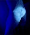
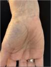
A 66-year-old woman presented with intense generalized itching after a trip abroad. Physical examination and manual dermoscopy confirmed the presence of multiple mite burrows (fig. 1) followed by a symmetrical papular eruption on the trunk and thighs. Subsequently, using a 365nm ultraviolet (UV) lamp, bright white-bluish structures corresponding to different parts of the mite burrow were seen, which supported the diagnosis of scabies (fig. 2).

Figure 1
(0.08MB).
Figure 2
(0.05MB).

The mite burrow is a well-known pathognomonic sign of scabies for dermatologists. Until now, visualization of the mite via optical microscopy, a technique known as Müller's test, is the diagnostic method of choice. However, this technique requires contact with the patient, training, and specific material to be performed. The 365nm UV LED light emits a white-bluish luminescence that is visible to the naked eye. When applied to the skin a bright white-bluish tunnel and a luminescent point corresponding to the mite's body can be seen. This technique seems to offer a new tool for diagnosing infestation by Sarcoptes scabiei and derivatives. Compared with Müller's test, this quick technique does not require direct contact, but a specific lamp.

Copyright © 2024. AEDV
Descargar PDF
Idiomas
Actas Dermo-Sifiliográficas
Opciones de artículo
Herramientas
es en

¿Es usted profesional sanitario apto para prescribir o dispensar medicamentos?

Are you a health professional able to prescribe or dispense drugs?