
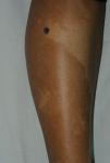

A 27-year-old woman, with no relevant medical or surgical history, consulted for an intensely pigmented papule that was located on the right leg, had appeared several years earlier, and had recently grown larger.
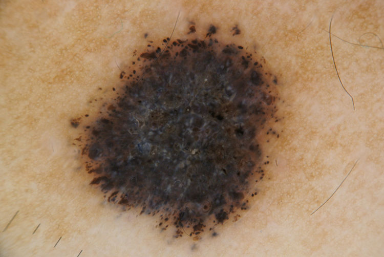
Physical ExaminationPhysical examination revealed a high phototype and a dark brownish black papule (8×9mm) (Fig. 1) on the lateral aspect of the right leg, lying on the border between a hypopigmented macule and normal skin. Dermoscopy revealed a central component with intense whitish blue–gray pigmentation, and the presence of evenly distributed dark-brown-to-black dots and globules in the periphery (Fig. 2).
Histopathology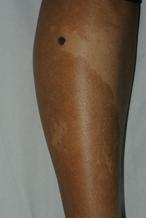
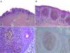
Biopsy of the excised lesion showed an acanthotic epidermis, the entire thickness of which contained markedly pigmented eosinophilic keratinocytes without atypia, no corneal plugs, mass necrosis and foci of differentiating cuticular cells, without involvement of or extension to the underlying dermis or signs of dysplasia or malignancy. Immunohistochemistry showed isolated cells positive for MelanA and HMB45 (Fig. 3).
![A, Acanthotic epidermis, the entire thickness of which contains nests of eosinophilic keratinocytes, without involvement of or extension to the dermis or underlying subcutaneous cellular tissue. Hematoxylin-eosin (H–E), original magnification ×10. B and C, Higher magnification images showing nests of markedly pigmented eosinophilic keratinocytes, without signs of atypia, absence of horny plugs, and mass necrosis and foci of differentiating cuticle cells (H–E, original magnification ×20 [B] and ×40 [C]). D, Image showing negative staining for HMB45 (original magnification ×20), despite the presence of intense pigmentation.](https://static.elsevier.es/multimedia/00017310/0000011300000003/v2_202212210529/S0001731022001314/v2_202212210529/en/main.assets/gr3.jpeg?xkr=ue/ImdikoIMrsJoerZ+w91sAmkCw32Jed9sZf6jzEuDbFpW7G0NfARZs8afh+9K8v8RN+oFy2ZmalFHXVYo6AgcJ0NBFNv9W/RaccXwyeDk+qBSBiG+kNAhHnXBjdH7fNWWcGbWHFuD0rld9zfzCI6e6OuSSdRFilNloBiKweio+zW9juoNOSirDQD/gfu105GhhDGu5bbOrfIxE7Fvi9GRG0Acf+bZ1ssTD+OCTBrdReAdITaosGh8V4K4BZnr8IF1kBJG9QpPAhQCAp4c5xsEglyeTWUGgH3z/qENDBLzXdRUH2FkztCgDsWwjne8W)
A, Acanthotic epidermis, the entire thickness of which contains nests of eosinophilic keratinocytes, without involvement of or extension to the dermis or underlying subcutaneous cellular tissue. Hematoxylin-eosin (H–E), original magnification ×10. B and C, Higher magnification images showing nests of markedly pigmented eosinophilic keratinocytes, without signs of atypia, absence of horny plugs, and mass necrosis and foci of differentiating cuticle cells (H–E, original magnification ×20 [B] and ×40 [C]). D, Image showing negative staining for HMB45 (original magnification ×20), despite the presence of intense pigmentation.
What Is Your Diagnosis?
DiagnosisPigmented hidroacanthoma simplex poroma.
CommentPoromas are a group of benign adnexal tumors that show terminal ductal differentiation of the sweat glands. Although historically this type of tumor has been considered to be of eccrine origin, there are probably similar numbers of eccrine and apocrine poromas.1 These usually manifest as solitary papules, plaques, or nodules that can appear in any location, although they show a preference for acral areas (palms and soles) and the scalp and, in the case of hidroacanthoma simplex, the extremities and trunk.2 They usually have a highly vascularized stroma, which lends a characteristic reddish pink color, although they can also be pigmented, as in the present case.
Definitive diagnosis is histological. Poromas are made up of 2 cell types: poroid cells, with a rounded basophilic nucleus and scarce cytoplasm, similar to the cells of the peripheral row of the distal portion of the duct; and larger, cuticular cells with scarce eosinophilic cytoplasm that resemble the luminal cells of the eccrine ducts.3 Depending on the architectural pattern, communication with the epidermis, and the presence or absence of solid and/or cystic components, 4 types of poromas can be distinguished: hidroacanthoma simplex, classic poroma, dermal duct tumor, and poroid hidradenoma. The degree of ductal differentiation varies, and therefore higher magnification can reveal the presence of sweat ducts inside the poroma, or dilation of the sweat ducts to form cystic spaces. Hidroacanthoma is limited to the epidermis, lacks communication with the dermis and tumor projections that exert pressure on the dermis (as occurs in the case of poroma), and has scant accompanying stroma.
It is characterized by abrupt demarcation between the lesion and the normal keratinocytes of the epidermis, which are more basophilic. This feature, and the absence of horny plugs, allows differentiation of hidroacanthoma from clonal seborrheic keratosis, which is the main differential diagnosis.2–4 Because this is a benign adnexal tumor, treatment is optional, and surgical removal is the treatment of choice.2
Shiiya et al.4 analyzed 4 cases of hidroacanthoma simplex and identified the presence of blood cells and fine black dots, fine annular scales, and the absence of glomerular vessels as characteristic findings of nonpigmented hidroacanthoma that are useful for differentiation from Bowen disease and seborrheic keratosis. In their study, Chessa et al.5 examined 26 histologically confirmed poromas, including 9 cases of hidroacanthoma simplex (4 nonpigmented and 5 pigmented). The pigmented lesions consisted of macules or slightly raised plaques, dermoscopic analysis of which revealed the presence of pseudo-reticulum, milium cysts, and comedone-like structures. None of the previously published dermoscopic descriptions of hidroacanthoma simplex reflect the features described in the present case.
In adnexal tumors, although dermoscopy can help orient the diagnosis,6 histopathology is essential for definitive diagnosis.


