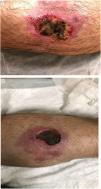
This is the case of 53-year-old man with a past medical history of malaria and schistosomiasis. The patient, a missionary in Benin, Africa, had developed a papular lesion on the posterior region of his right lower limb upon his return. During the initial physical examination, no significant signs or symptoms were found. Initially, at a different hospital, rickettsiosis had been suspected based on positive serology, and treatment with doxycycline, along with surgical debridement had been initiated, but remained unresponsive. Over time, the lesion had progressed to a skin ulcer (Fig. 1A).
In our hospital, the patient was reevaluated, and a biopsy culture was performed using Löwenstein-Jensen medium, and a PCR assay for M. ulcerans detection and histopathology, both of which turned out initially negative. Given the clinical presentation and the patient's origin (Benin), Buruli ulcer was suspected.
An 8-week course of empirical treatment with rifampicin 600mg/day plus moxifloxacin 400mg/day was started, with favorable outcomes (Fig. 1B).
Diagnostic confirmation was based on the patient's good response to the initiated treatment, and the presence of AFB in Löwenstein cultures at 9 months. However, a PCR assay could not be performed because of the small amount of AFB. Attempts to close the ulcer with 2 grafts failed due to overinfection. Currently, the patient is undergoing local wound care with slow wound healing.


