


Syphilis —the “great simulator” for classical venereologists—is re-emerging in Western countries despite adequate treatment; several contributing factors have been identified, including changes in sexual behaviour, which won’t be the topic of this article though.
In 2021, a total of 6613 new cases of syphilis were reported in Spain, representing an incidence of 13.9×100 000 inhabitants (90.5%, men). Rates have increased progressively since 2000.
The clinical presentation of syphilis is heterogeneous. Although chancroid, syphilitic roseola and syphilitic nails are typical lesions, other forms of the disease can be present such as non-ulcerative primary lesions like Follmann balanitis, chancres in the oral cavity, patchy secondary lingual lesions, or enanthema on the palate and uvula, among many others.
Regarding diagnosis, molecular assays such as PCR have been replacing dark-field microscopy in ulcerative lesions while automated treponemal tests (EIA, CLIA) are being used in serological tests, along with classical tests (such as RPR and HAART) for confirmation and follow-up purposes. The interpretation of these tests should be assessed in the epidemiological and clinical context of the patient. HIV serology and STI screening should be requested for anyone with syphilis.
Follow-up of patients under treatment is important to ensure healing and detect reinfection. Serological response to treatment should be assessed with the same non-treponemal test (RPR/VDRL); 3-, 6-, 12-, and 24-month follow-up is a common practice in people living with HIV (PLHIV).
Sexual contacts should be assessed and treated as appropriate.
Screening is advised for pregnant women within the first trimester of pregnancy. Pregnant women with an abortion after week 20 should all be tested for syphilis.
The treatment of choice for all forms of syphilis, including pregnant women and PLHIV, is penicillin. Macrolides are ill-advised because of potential resistance.
La sífilis, la «gran simuladora» de los venereólogos clásicos, está resurgiendo en países occidentales a pesar de existir tratamiento adecuado; diversos factores contribuyen, entre ellos cambios de comportamientos sexuales, no siendo objeto de este trabajo describirlos.
En 2021 en España se notificaron 6.613 nuevos casos que representan una incidencia de 13,9×100.000 habitantes, 90,5% varones. Las tasas han aumentado progresivamente desde el año 2000.
La presentación clínica es heterogénea. Aunque el chancro, la roséola sifilítica y los clavos sifilíticos son lesiones típicas; destacamos otras formas, como las lesiones primarias no ulcerativas como la balanitis de Follmann, los chancros, en cavidad oral, las lesiones secundarias linguales parcheadas o el enantema en paladar y úvula, entre muchas otras.
Respecto al diagnóstico, las técnicas moleculares PCR están desplazando al campo oscuro en lesiones ulcerativas y en el análisis serológico se emplean pruebas automatizadas treponémicas (EIA, CLIA) que se combinan con pruebas clásicas (como RPR y TPHA) para la confirmación y el seguimiento. La interpretación de estos test debe valorarse en el contexto epidemiológico y clínico del paciente. Se debe solicitar serología de VIH y cribado de infección de transmisión sexual a toda persona con sífilis.
Es importante realizar un seguimiento de los pacientes tratados para garantizar la curación y detectar reinfecciones. Se aconseja valorar la respuesta serológica al tratamiento con la misma prueba no treponémica (RPR/VDRL) cuantificada. El seguimiento de los controles se realiza a los 3, 6 y 12 meses extendiendo a 24 en las personas viviendo con VIH (PVV).
Los contactos sexuales deben ser evaluados y tratados según proceda.
Se recomienda el cribado en embarazadas en el primer trimestre de gestación. Toda mujer con aborto de más de 20 semanas debe ser testada de sífilis.
El tratamiento de primera elección en todas sus formas, incluso embarazadas y PVV, sigue siendo la penicilina. Los macrólidos no se recomiendan dada la potencial resistencia.
Syphilis is a sexually transmitted infection (STI) considered a notifiable disease (ND) in all Spanish autonomous communities.1 Syphilis is known as “the great imitator” because the lesions it causes can be confused with those of multiple diseases.
EpidemiologyThe prevalence of syphilis is high in low- and middle-income countries, although its incidence in high-income countries has been on the rise over the past 25 years, mainly among men who have sex with men (MSM). There is an increased incidence associated with HIV infection, unprotected sex, and in some countries, with the recent implementation of pre-exposure prophylaxis (PrEP) for HIV prevention.2
In 2021 in Spain, a total of 6613 new cases of syphilis were reported (an incidence of 13.97/100,000 inhabitants). The lowest rates were reported in the year 2000 in the period that goes from 1995 through 2021. Since then, they have gradually increased to reach historic peaks in 2021. A total of 90.5% of all reported cases were men (86.6% in MSM). The median age was 36 years, with no differences being reported by sex, and the highest rates (41.62/100,000) being reported in the 25 to 34-year-old age group. Only 33.9% provided data on HIV co-infection, and 9.21% of these were people living with HIV (PLHIV).
Etiology and transmissionSyphilis is caused by Treponema pallidum subsp. pallidum, a 6-20nm x 0.1-0.18nm gram-negative bacterium (called pallidum because of its poor affinity for gram staining) of the order Spirochaetales. Its small size does not make it visible with conventional optical microscopy. Furthermore, it is non-cultivable and moves with a characteristic corkscrew motion due to its endoflagella.3 It is an obligate parasite, and humans are its only reservoir.
It is transmitted through direct contact with an infectious lesion on affected skin or mucous membranes (mainly through sexual contact), by blood, and transplacentally.3 When transmitted through direct contact, the bacterium penetrates through small erosions on the skin reaching the dermis and subcutaneous tissue, where it multiplies evading the innate immune response and spreading lymphatically and hematogenously, reaching the remaining tissues.
The presence of syphilitic mucocutaneous lesions promotes the transmission of HIV.2
Clinical signsEarly syphilisPrimary syphilisAlso called chancre, it appears at the inoculation site after an incubation period of 10-90 days.4 Although traditionally described as a single, painless, indurated, reddish ulcer of 0.5cm to 3cm in diameter (fig. 1),4,5 a study confirmed that it can sometimes be painful (49.2%) or multiple ulcers.6 It is usually associated with a loco-regional adenopathy7 and resolves within 3-6 weeks without scarring if left untreated.8

Although it is generally located in the anogenital region, it can appear in any exposed areas, including mouth, fingers, nipples, etc.9.
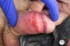
Syphilitic balanitis of Follmann is a less common, possibly underdiagnosed presentation (fig. 2), presenting as erosive and painful balanitis.6,10
Secondary syphilis
The hematogenous and lymphatic dissemination of spirochetes occurs 3 to 12 weeks after the resolution of the chancre (although both stages may overlap) and results in a wide array of clinical signs.
Mucocutaneous signs are the most common ones—in up to 97% of patients4—and are usually accompanied by systemic signs and symptoms, such as generalized lymphadenopathy, malaise, sore throat, myalgia, headache, and low-grade fever4.
We call syphilides11 to all those mucocutaneous signs of early syphilis other than chancre, which can be localized or generalized and are generally mildly symptomatic.
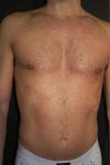
The most common presentation is a diffuse maculopapular rash on the trunk and extremities with fine scaling called “roseola” (fig. 3). Numerous atypical forms of cutaneous presentation have been reported, such as nodular, pustular, lichenoid, psoriasiform, annular, follicular, ulceronodular (also called malignant syphilis, etc.)12,13. Malignant syphilis is a rare and aggressive presentation consisting of necrotic ulcers and nodules. It is associated with HIV infection, low CD4 count, malnutrition, MSM, previous syphilis, diabetes mellitus, and alcohol abuse14.

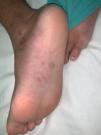
Syphilides may appear on palms and soles in up to 40%-80%4 of cases, often exhibiting reddish-brown macules with or without a slight collarette of scaling called clavos (fig. 4).

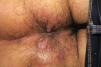
Lesions in the anogenital region are a common finding, appearing as patches or geographic lesions (fig. 5). In areas prone to maceration, exophytic, moist, and friable lesions called condylomata lata may appear (fig. 6).4 They can be confused with condylomata acuminata and tumor lesions7, and are highly contagious.


Oral mucosal involvement occurs in up to 30%-40%15 of patients, and these lesions are also highly infectious. Small, rounded patches on the dorsal tongue, larger depapillated plaques, and erosions on the tongue or lips are common findings (fig. 7).15 Other oral presentations include “rhagades,” enanthema, and whitish plaques on the palate, uvula, and tonsils (fig. 8).16


Alopecia is a less common sign, and is usually of moth-eaten appearance (fig. 9)15.

If left untreated, secondary syphilis usually resolves spontaneously within a matter of 4 to 12 weeks without leaving a scar.
Early latent syphilis or early non-primary non-secondary syphilisThe term early latent syphilis describes patients without any signs or symptoms of primary or secondary syphilis, but with positive serologic tests and evidence that the infection was acquired over the past 12 months.5,17,18
Late latent syphilis (or of unknown duration)Late latent syphilis refers to infections without any signs or symptoms of syphilis and no indications of contagion over the past 12 months, only the serologic evidence of infection (or reinfection).18
These patients should undergo a thorough examination to evaluate the possible presence of lesions (primary or secondary syphilis).
A small percentage of untreated syphilitic patients will develop clinical signs years after the infection.4 Cardiovascular syphilis and gummatous syphilis are currently rare, yet neurosyphilis is more prevalent.19
Neurosyphilis, ocular syphilis, and otosyphilisAlthough neurosyphilis, otosyphilis, and ocular syphilis can occur at any stage of the infection, these are not stages of the disease per se. Neurosyphilis can be asymptomatic (evidence of central nervous system infection without clinical signs). Progression into symptomatic neurosyphilis is extraordinarily rare,20 so lumbar puncture (LP) is ill-advised in most asymptomatic patients.5
The most common symptoms of early neurosyphilis are mild meningeal signs, such as headache and nausea. Neurosyphilis can cause cranial nerve paralysis or meningovascular involvement.
Late symptomatic neurosyphilis is much less common,4 causing general paresis (paralytic dementia) and tabes dorsalis.
Ophthalmic signs of syphilis are varied, such as red eye, blurred vision, vision loss, etc. They frequently appear during the secondary stage of the disease and can affect any segment of the eyeball21. The most common diagnosis is uveitis.21
Otosyphilis is a rare inner ear disease presenting as unilateral or bilateral hearing loss, tinnitus, or vestibular disturbances, which can be reversed if treated early.22
Congenital syphilisTreponema pallidum infection can occur in the fetus of any untreated infected mother. It is most likely within the first year after acquiring the disease (85%-90% of cases)23 in immunocompromised patients, and after 16-20 weeks of pregnancy.23 Infection during delivery is also possible.
If left untreated, fetal/neonatal death occurs in 40% of cases, while in the remaining 60%, two-thirds will be asymptomatic at birth.23
Symptoms of congenital syphilis- -
Early (< 2 years): mucocutaneous syphilides, palmoplantar pemphigus, rhinitis, jaundice, lymphadenopathies, meningitis, nephrotic syndrome, hemolytic anemia, prematurity, bone lesions, etc.23,24.
- -
Late (> 2 years): deafness, interstitial keratitis, dental anomalies, bone lesions, neurological or gummatous involvement, etc.23.
Treponema pallidum is not cultivable in laboratory media, so direct diagnosis is based on detecting it in ulcerated or exudative lesions through dark-field microscopy, which can identify its morphology and motility.25,26 Although this method can be useful for genital ulcers with negative serological screening in centers with a significant volume of samples and experienced microscopists,27 a negative result does not exclude the disease.
Polymerase chain reaction (PCR)This is currently the most widely used technique for direct diagnosis. It is the method of choice for ulcerated or erosive oral, anal, and other exudative lesions where commensal treponemas exist. The PCR is also useful in the newborns’ vitreous humor, placenta, and exudative tissues; however, it has low sensitivity in cerebrospinal fluid (CSF),28 and its yield varies depending on the type of sample and the stage of the infection, being high in primary ulcerative lesions and lower in secondary lesions.29
Commercially available multiple platforms detect different agents causing ulcerative STIs.
Direct immunofluorescence techniques, in situ hybridization, or silver staining techniques are currently not used anymore.30,31
Serological techniquesNon-treponemal tests (Ntts)Serological diagnosis is indirect and presumptive, not differentiating among different pathogenic treponemas (T. pertenue, T. endemicum, and T. carateum) .32
Non-treponemal or reagin tests use antigens composed of cardiolipin, lecithin, and cholesterol and are primarily the Rapid Plasma Reagin (RPR) and Venereal Disease Research Laboratory (VDRL) tests. Both are manual, simple, inexpensive, and semi-quantitative techniques to assess disease activity and post-treatment monitoring. They test positive 10-15 days after the appearance of the chancre if left untreated. Titers peak 1 and 2 years after infection and remain low positive in late untreated disease.26 A quantified serum sample should be obtained before treatment (or within the first few hours) to have a baseline test and measure subsequent changes with the same technique (1, A). Ntts are quantified as follows: 1/1 (pure serum), 1/2, 1 /4, 1/8, 1/16, 1/32, 1/64, etc.
Seroreversion is a 4-fold decrease of titers (2 dilutions) between 6 and 12 months after early infection (e.g., from 1/16 to 1/4) and indicates adequate treatment.33 Occasionally, some patients properly treated based on their stage fail to reduce Ntt titers by 4 times (at least, 2 dilutions) at the 6-to-12-month follow-up for early syphilis and at the 12-to-24-month follow-up for late syphilis in the absence of reinfections; this lack of response is called serofast reaction and is influenced by factors, such as the stage of the disease, duration, and initial Ntt titer. Its causes are not entirely clear.34–36 We should think of reinfections or relapses (treatment failures) when Ntt titers increase by 4 times or 2 dilutions after correct treatment.
Ntts can show false positive in 0.2% up to 0.8% of cases and less frequently in treponemal tests (see annexes 1 and 2, supplementary data).37
Treponemal tests (TTs)Treponemal tests are qualitative and earlier than Ntts. They detect specific antibodies 2 to 4 weeks after exposure.32 They are used as confirmatory tests and are not useful to monitor treatment or disease activity as they remain positive in most treated cases.26 The most widely used are T. pallidum hemagglutination (TPHA), T. pallidum microhemagglutination (TP-MHA), fluorescent treponemal antibody absorption (FTA-ABS), IgG or IgM immunoblot, enzyme immunoassay (EIA), and chemiluminescence immunoassay (CLIA).
EIA and CLIA tests are automated and allow testing sera from multiple patients, making them a crucial screening tool.
Although false positive TTs are possible, they are less frequent than Ntts (see annexes 1 and 2).37
Most laboratories use the so-called reverse algorithm38 as a screening test, performing automated EIA or CLIA (both TTs), which are the most efficient; positive tests may be due to past treated disease or an untreated patient with active disease. An initial positive test should be confirmed with another TT, usually TPHA (1, C); if positive, a quantified Ntt should be performed before establishing the baseline titer, which indicates activity and serves as post-treatment control (1, A).
The clinical and epidemiological context should always be considered when interpreting syphilis tests (annex 3).39
NeurosyphilisCSF evaluation is ill-advised in early syphilis in patients without neurological, ocular, or auditory symptoms (1, A). It is, however, indicated in patients with neurological symptoms,40 regardless of the stage of the disease (1, C), and in syphilis with ocular involvement, it should be individually assessed.36
CSF examination includes total proteins, the number of mononuclear cells, treponemal tests (FTA or TPHA), and non-treponemal tests, preferably VDRL.
No single test per se can confirm the presence of neurosyphilis. While a positive VDRL test in CSF is considered diagnostic of late-stage neurosyphilis in the absence of blood contamination, a negative result does not exclude diagnosis.17,41 PCR in CSF has low sensitivity and specificity rates for neurosyphilis diagnosis.28
Neurosyphilis diagnosis is rare in patients with negative blood Ntts (data provided in the presentation “Syphilis & neurosyphilis update” at the IUSTI 2023 Congress held in Malta, Dr. Nicolas Dupin, Professor of Dermato-Venerology at University Paris Cité, Cochin Hospital, APHP. Head of the National Reference Center of Syphilis, Former president of the French Society of Dermatology).
TreatmentPrimary, secondary, or early latent syphilis5,17,18First-line therapyBenzathine penicillin G (BPG) 2.4 million international units (MIU) intramuscular (IM) (1, B).
If allergic to penicillin, if parenteral treatment is refused, or in the presence of bleeding disorders: doxycycline 100mg orally every 12hours for 14 days (1, C).
Azithromycin is ill-advised due to the potential resistance of Treponema pallidum.28,42–44
Late latent syphilis or of unknown duration, cardiovascular or gummatous involvement5,17,18First-line therapyBenzathine penicillin G 2.4 MIU IM, weekly dose for 3 weeks (1, C).
If allergic to penicillin, if parenteral treatment is refused, or in the presence of bleeding disorders: doxycycline 100mg orally every 12hours for 4 weeks (2, D).
Neurosyphilis, ophthalmic, and otic involvement5,17,18First-line therapySodium penicillin G (also known as benzylpenicillin) 3-4 MIU IV every 4hours for 14 days (1, C) or 18-24 MIU/day in continuous IV infusion for 14 days.
Alternatives: IV ceftriaxone 2g daily for 10-14 days (1, C); procaine penicillin 2.4 MIU IM daily plus probenecid 500mg every 6hours for 10-14 days (1, C).
Penicillin allergyDesensitization and subsequent treatment with penicillin as the first-line therapy is recommended (1, C). The duration of the recommended and alternative regimens in neurosyphilis is shorter than treatments for latent syphilis, which is why some reports consider additional doses of benzathine penicillin 2.4 MIU IM weekly for 3 weeks after the IV treatment, providing a therapeutic duration comparable to latent forms.18,45
Summary of therapy inTable 1
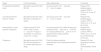
Therapeutic recommendations for syphilis treatmenta
| Stage | First-line therapy | Other alternatives | Comments |
|---|---|---|---|
| PrimarySecondaryEarly latent | Benzathine penicillin G2.4 MIU IM | Oral doxycycline 100mg every 12hours, 2 weeks | Request HIV testSerological controls: RPR or VDRL at 3, 6, and 12 months |
| Late latentUnknown duration Tertiary | Benzathine penicillin G2.4 MIU IM once weekly for 3 weeks | Oral doxycycline 100mg every 12hours, 4 weeks | Request HIV testSerological controls: RPR or VDRL at 3, 6, 12, and 24 months |
| Neurosyphilis, ocular syphilis, otosyphilis | Sodium penicillin G3-4 million units IV every 4hours for 14 days or 18-24 MIU on continuous infusion for 14 days | Procaine penicillin G2.4 MIU IM daily plus probenecid 500mg every 6hours for 10-14 daysCeftriaxone 2g IV for 10-14 daysPenicillin-allergic patients: desensitization | Request HIV testSerological controls: RPR or VDRL at 3, 6, 12, and 24 monthsPeriodic CSF exam |
| Pregnancy | Benzathine penicillin G2.4 MIU IM once weekly for 1 to 3 weeks depending on stage | Penicillin-allergic patients: desensitization and subsequent penicillin treatment | Request HIV test Serological controls: RPR or VDRLObstetric follow-up |
The recommendations stated in this article may not be appropriate for use in all clinical situations. Decisions to follow these recommendations should be based on the physician's best professional judgment and consideration of the individual circumstances of each patient and available resources.
Treatment for HIV-positive patients should be administered the same as for non-HIV-infected patients, with careful monitoring to ensure an adequate response.5
b Some clinical practice guidelines17,44 propose completing treatment with benzathine penicillin G 2.4 MIU IM once a week for 3 weeks after IV treatment.
Source: Janier et al.5, Kingston et al.17, and Workowski et al.18.
All individuals diagnosed with syphilis are recommended to undergo clinical and serological evaluation 3, 6, and 12 months after treatment18 (1, D). Their serological responses should be compared with the titers of the same Ntt (RPR/VDRL) obtained on the same day of treatment,5,18 or as close to this date as possible. HIV serology and screening for other STIs should be requested. If the risk of reinfection is high, frequent Ntt checks (e.g., every 3 months) are advised (2, C).5
A negative Ntt after treatment is considered the best confirmation of cure, although it is not achieved in all cases.
Reinfection or therapeutic failure should be considered if a person maintains signs or symptoms, if these reappear, or if there is an increase of, at least, 4 times the titer (2 or more dilutions) of the Ntt remaining elevated for more than 2 weeks.5,18,46
An increase in Ntt in sexually active individuals correctly treated and without neurological symptoms would more likely indicate reinfection rather than therapeutic failure, so it is recommended to re-treat based on staging (1, C), repeat HIV serology,18 and re-evaluate contacts.5
If after 6-12 months of treatment there is no 4-fold decrease in Ntt (“serological failure”), some professionals recommend additional treatment with a 3-week regimen of a weekly injection of benzathine penicillin G 2.4 MIU (unless there are neurological symptoms or CSF abnormalities), although there is no solid evidence for this recommendation (2, D).5
In the presence of neurological symptoms, a CSF exam is necessary regardless of the stage of the disease (1, C).47
Despite correct therapy and a negative CSF exam, serological titers may not decrease. In these cases, retreatment or CSF exam is not recommended.34
Up to 10%-20% of individuals treated according to recommendations may not achieve a 4-fold decrease in titers within a year.48,49 Numerous factors are associated with the serological response, such as staging (in early stages a 4-fold decrease in titers is more likely), initial Ntt titers (levels <1/8 respond worse vs higher levels), and age (younger individuals achieve the 4-fold decrease vs older individuals),50 syphilis reinfections (higher titers with slower decrease).18,51 If therapeutic failure without sexual relations in the past 3-6 months is suspected, with the possibility of asymptomatic neurosyphilis (low evidence), some authors recommend performing a CSF exam, repeat HIV serology, and findings-based treatment.5
In late latent forms, Ntt titers are usually negative. In individuals not living with HIV, with adequately treated late latent syphilis and low but stable Ntt titers, follow-up is not required (2, D).5
It is recommended to repeat the CSF exam 6 weeks to 6 months after neurosyphilis treatment to see the decrease in proteins and white cells (2, D). This exam could be avoided if Ntt negativize (2, D).52
Management of special populationsPeople living with HIV (PLHIV)PLHIV should be treated with the same guidelines as the rest of the population (1, B)5,17,18. Closer monitoring can be recommended if CD4 levels are <350/mm3 or if they are not on antiretroviral treatment (2, D).
PregnancyEvery woman should undergo syphilis serology testing at the first prenatal visit (1, A). A non-treponemal titer > 1/8 may be indicative of early active infection. Women living in communities with high syphilis rates (rates > 7.73 cases/100,000 inhabitants)53 or at high risk of infection are recommended to undergo serological follow-ups within the third trimester (28 weeks) and at deliver18,54. Additionally, any woman with a miscarriage after week 20 should be tested for syphilis.18 No mother or newborn should be discharged without the mother being tested for syphilis, at least, once during pregnancy.
The risk of vertical transmission depends on the stage of syphilis during pregnancy, being higher in primary and secondary stages, and lower in late stages of the disease with low titers. Pregnant women with low and stable titers previously treated do not require new treatment unless there is an increase in these titers (> 2 dilutions), indicating possible reinfection or treatment failure.
The only accepted treatment during pregnancy is penicillin, using the recommended regimen according to the stage of syphilis. However, some sources recommend an additional dose of 2.4 MIU of benzathine penicillin G 1 week after the initial treatment (1, B) for pregnant women diagnosed during the primary, secondary, or early latent stages of the disease.54,55
Diagnoses of syphilis during the second half of pregnancy require fetal ultrasound monitoring. If infection-related abnormalities such as hepatomegaly, ascites, placental thickening, etc. indicating a higher risk of treatment failure are found, a second dose of penicillin 1 week after the first one is even more justified.18 In late latent stages of the disease requiring 3 doses, subsequent doses should not be delayed more than 9 days.
Pregnant women allergic to penicillin should be desensitized and treated with benzathine penicillin G (1, C).5,17,18
Before treatment, patients should be informed of a possible Jarisch-Herxheimer reaction, which in the second half of pregnancy could induce preterm labor.54 Pregnant women should be evaluated by an obstetrician if they experience fever, contractions, or decreased fetal movement after treatment.
Contact managementAll sexual contacts of a person diagnosed with primary, secondary, or early latent syphilis should be clinically and serologically evaluated and treated as appropriate, following these recommendations5,17,18:
- a.
Sexual contacts within 90 days prior to syphilis diagnosis; treat as early syphilis, even if serology is negative18.
- b.
Sexual contacts > 90 days prior; treat as early syphilis if serological testing is not immediately available or if follow-up of the contact is uncertain. If serology is negative, no treatment is needed. If positive, act according to clinical presentation, serology, and stage of syphilis18.
- c.
Sexual partners with ongoing contact with patients with late latent syphilis should be clinically and serologically evaluated for syphilis and properly treated.5,17,18
- d.
Follow-up is necessary for at-risk contacts, including partners who had sexual contact more than 3 months ago with someone diagnosed with primary syphilis, more than 6 months ago with someone diagnosed with secondary syphilis, and 1 year ago with someone diagnosed with early latent syphilis.5
None declared.


