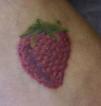
A woman aged 23 years, with no past medical history of interest, was seen for the appearance a week earlier of pruritic skin lesions in the area of a tattoo created on the dorsum of her right foot 6 months earlier (Fig. 1). The patient denied taking any new medication or applying any topical products. There were no lesions on the rest of her skin or mucosas or lesions of the skin appendages. Dermatologic examination revealed a shiny elevated plaque in the area of the tattoo, producing a 3D morphological pattern of a strawberry, with minute coalescent whitish papules predominantly affecting the borders of the tattoo. Histology was compatible with a lichenoid reaction. Treatment was started with 0.5% clobetasol propionate cream daily for 2 weeks, with no improvement.
Tattoos can give rise to various complications, including acute or chronic infectious conditions, benign or malignant tumors, dermatoses induced by a Koebner phenomenon, and acute or chronic inflammatory changes with a variety of histological patterns. Lichenoid reaction to the red dye of tattoos is the most common chronic inflammatory disorder. The pigment is based on mercury, which is the most frequently implicated etiological factor.
Please cite this article as: Imbernón-Moya A, Fernández-Cogolludo E, Gallego-Valdés MÁ. Tatuaje de fresa en 3D. Actas Dermosifiliogr. 2017;108:950.


