In 1936, Covisa and Bejarano published a treatise entitled Elementos de Dermatología (The Elements of Dermatology). In this surprisingly modern book they abandoned the nosological debates characteristic of the 19th century and instead classified diseases according to their etiology and pathogenesis based on the scientific and technical advances of the time. Moreover, unlike other books available at the time, which were essentially adaptations of foreign texts, this was the first medical work to reflect the reality of Spanish medicine.
However, the future of both the book and its authors was to be determined by the start of the Spanish Civil War in the same year. Covisa and Bejarano, who were both extremely active in the public health system and medical education during the Second Republic, were obliged to seek exile in America. Due to the difficulties of the time, very few copies of the book reached the public and no new editions were ever printed. We will never know what would have happened if the war had not started, but we believe that this important work should be remembered.
En 1936 Covisa y Bejarano publicaron su tratado titulado Elementos de Dermatología. El libro destacaba por su modernidad, al dejar atrás los debates nosológicos que caracterizaban a la Dermatología del siglo anterior, y al agrupar las enfermedades por su etiología y patogenia, apoyándose en los avances científicos y técnicos de la época. Era también el primer texto adaptado a la realidad española, y no una simple adaptación de un texto extranjero.
Sin embargo, la Guerra Civil iniciada ese mismo año determinó el futuro de los autores y del propio libro. Covisa y Bejarano tuvieron una intensa participación en la administración sanitaria y universitaria de la Segunda República y se vieron obligados a exiliarse a América. El libro tuvo escasa distribución por librerías en aquel difícil momento, y tampoco se realizarían nuevas ediciones. Nunca sabremos qué habría sucedido de no haber estallado la guerra, pero creemos justo recordar esta importante obra.
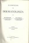
Sánchez-Covisa and Bejarano's Elements of Dermatology1 (Fig. 1) was not a dense treatise for specialists only, as its very title showed. The book mainly targeted student readers and general practitioners, for whom the authors sought to explain the fundamental concepts of skin diseases that would be needed for competent clinical practice. As followers of Juan de Azúa, who never managed to publish his own general textbook, the authors saw this book as a way to collect their mentor's teachings and ensure continuity.
This was not just another textbook, however. It was the first modern account of dermatology by Spanish authors.
Sánchez-Covisa and Bejarano organized the content mainly around the causes of disease, the mechanisms of pathogenesis. In this way, they avoided becoming entangled in the morphologic classifications and assumptions about predispositions and imbalanced humors that were so characteristic of earlier treatises on the subject.
Furthermore, instead of taking a foreign text as their starting point, these authors selected the skin conditions that were most common in Spain and whenever possible they cited Spanish sources.
Because of the Spanish Civil War, however, and the persecution and purges that followed it under the regime of Francisco Franco, any vestiges of the Republic that remained were erased. This textbook and its authors therefore never gained the recognition they deserved.
Historical Context for the Publication of The Elements of DermatologyIn addition to their work as scientists and educators, Sánchez-Covisa and Bejarano engaged intensely with politics, and that activity would mark their lives in the years following the book's publication. Politics would also affect the future of the book itself. To understand the timing of publication, it will be sufficient to look at the page on which the authors dedicated their work to Professor de Azua. The date was January 1936, and the Spanish Civil War would start 6 months later.
Sánchez-Covisa declared himself an agnostic and liberal—and a supporter of the Republic. In Cuenca, where he had been born 50 years earlier, he stood for election to the 1931 constitutional convention (Cortes Constituyentes) as a member of the party founded the year before by Niceto Alcalá-Zamora (the Liberal Republican Right). Disagreements over ideology led him to quit that party the same year, and in 1932 he joined Manuel Azaña's Republican Action party.2 He ceased to be a parliamentarian in 1933 when the constitutional convention was dissolved. Sánchez-Covisa had been named professor of dermatology and syphilology at Madrid's central university in 1926, succeeding de Azua after a selection process open to all interested candidates.3,4 Later, during the Second Republic, he became dean5 and held that position until 1934. He also served as president of Madrid's College of Physicians and of the Academy of Physicians and Surgeons, as minister of health,2 and of course as president of the Spanish Academy of Dermatology and Venereology (AEDV) and editor of Actas Dermo-Sifiliográficas.6
The military insurrection led by General Franco in 1936 triggered the Spanish Civil War. In August of the same year, Sánchez-Covisa was named head of the university's Hospital Clínico by order of the education ministry of the Popular Front,7 with which he announced his affiliation at his first meeting with the hospital's staff.2,8 In November 1936 he moved to Valencia with the government without prior warning, attracting criticism.8 He later went to Barcelona and, after the war, into exile. Sánchez-Covisa had taken an active part in the government of the Second Republic and was subject to persecution by Franco's nationalist supporters, yet his own side considered him too much of a moderate. After going first to Paris and then to New York, Sánchez-Covisa accepted the invitation of the Ministry of Health and Social Welfare of Venezuela. There he served as an advisor to the ministry's venereology division.9,10 When he died in Caracas on June 24, 1944, he was seeking permission to go to Portugal.2
Bejarano was initially Sánchez-Covisa's disciple but soon became his colleague and close collaborator, even sharing with him the leadership of the dermatology department at Hospital San Juan de Dios. He served as president of the AEDV and presided over the events celebrating the society's 25th anniversary in 1934.3,6 In 1933 he was named director general of health by the Republican government but remained in office only 4 months.11,12 When the Popular Front took charge of the College of Physicians in July 1936, he became president of that body.13 Later Negrín made him head of health services at the military police's training facility (Instituto de Carabineros).14 Bejarano also moved to Valencia with the government during the war and finally went into exile in Mexico, where he was elected president of the Mexican Society of Dermatology and directed a leprosy hospital in Zoquiapan, near Puebla.15 He died in exile in 1965.3
Serviliano Pineda, who produced the photomicrographs for the textbook, was probably active in Communist Party circles and was a member of the Popular Front Committee at the Hospital Clínico during the war; in the purges that followed, the university's political tribunal stripped him of appointments and declared him unfit for posts requiring public trust or the exercise of authority.8,15
Dermatology, From Morphology to EtiologyThe 20th century began with marked technological advances. In dermatology in particular, discoveries in microbiology and pathology led the discipline to question doctrines inherited from the previous century.
Pierre Antoine Ernest Bazin, preeminent exponent of the French school of dermatology during the second half of the 19th century, argued the theory of diathesis, or the existence of constitutions that predisposed an individual to certain skin diseases. In Spain, José Eugenio Olavide subscribed to this view. In contrast, the Vienna school, led by Ferdinand Ritter von Hebra, favored concepts from pathology, classifying dermatoses based on symptoms and local factors. The theories of the Viennese school were the ones de Azúa adopted. Sánchez-Covisa and Bejarano, as contemporaries of Paul Gerson Unna, would also have witnessed the emergence of the Hamburg school, which drew on the disciplines of pathology, chemistry, and microbiology.
As the technical means to investigate the etiology and pathogenesis of disease had become available, professionals had come to the conclusion that classifications based on diathetic concepts or purely morphological observations were inexact and impractical. Not all dermatoses could be classified according to the new criteria, however, because their causes were still unknown—just as some remain unknown today. Louis Anne Jean Brocq, another contemporary of Sánchez-Covisa and Bejarano, attempted an etiologic classification of skin diseases of known cause, drawing on the theory of diathesis to a certain degree.
The authors of The Elements of Dermatology attempted to impose order by distinguishing between dermatoses of known, unknown, and multifactorial causes, along the lines of Brocq. However, they used other criteria as well, for example grouping diseases by adnexal location or according to the special natures of certain conditions, such as tumors. At the time, these ideas were revolutionary.
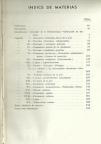
The Physical Characteristics of the BookThe textbook is bound between board covered with dark blue cloth embossed with gold lettering. The front matter (including a table of contents, dedication and foreword) takes up 11 pages, the main body comprises 547 pages of content, and a page at the end lists errata. The writing is clear and practical, with marginal notes at the beginning of each section. The names of cited authors appear in capital letters.
An interesting feature is the liberal use of illustrations, of which there are 253 original figures in black and white. All were derived from cases treated within the authors’ own practice and were prepared under their supervision. Among them are both clinical photographs and photomicrographs from the university department's pathology laboratory. The images were of such good quality for the period that it was not considered necessary to provide an additional sketch or outline by way of explanation.
In their preface, the authors wrote, “We hope this work has a national character to the extent that is possible. By this we mean that it reflects the peculiar nature of skin diseases in our country, their frequency, clinical presentations, and therapeutic indications.” In fact, the scope of subjects covered differs from those that would be included today. Infectious diseases carried more weight then, and particular attention was given to leprosy and tuberculosis (Fig. 2).
The Scope of ContentAlthough the following 4 categories are not named as subdivisions in the table of contents, they provide a convenient way to conceptualize the scope of the book.
General Introductory ChaptersGeneral aspects of the anatomy, histology, and physiology of healthy skin are treated in the first 5 chapters, along with the general etiology, pathogenesis, diagnosis, and treatment of skin diseases.
These first chapters reveal how different treatments were when this book was published in comparison with today's therapeutic arsenal. Active agents were few, but they included such products as gold salts, sulfates, reducers, arsenic compounds, and chrysarobin. For topical application, the main concern was to find the most appropriate formulation, choosing among powders, water-based or other pastes, oils, ointments, or plasters. Dietary interventions, such as the Gerson diet for tuberculosis, were prescribed. Autohemotherapy, vaccination, and desensitization were also used. However, we must take note of the role played by physical modalities, particularly roentgen (x-ray) therapy, which was used for psoriasis, chronic eczema, lichen planus, ringworm, postherpetic neuralgia, skin tumors, lupus erythematosus, and tuberculosis. X-rays were applied to the skin directly or indirectly targeted sympathetic nerves or the spleen. Bucky and radium rays were also used, along with electrotherapy, dry ice, high-frequency currents, and phototherapy (mainly with the Finsen light).
Exudative and Erythematosquamous DermatosesThe first group of diseases covers exudative and erythematosquamous dermatoses: eczemas, psoriasis, seborrheic dermatitis, pemphigus and other so-called pemphigoid conditions, forms of erythema multiforme (including erythema nodosum), and herpes. References to the work of Bejarano and Gómez Orbaneja on the ties between psoriasis and rheumatic conditions stand out in these chapters. The authors note that these ties are “still obscure and difficult to interpret, but the problem is surely a very general one with a wide scope since it has led very different processes to become analogous in so many details.” They later discuss “epidemiological, clinical, and anatomic relationships between herpes zoster and chicken pox,” a subject that was carefully studied by Sáinz de Aja.
Another interesting section deals with alopecias, in the chapter on seborrhea and seborrheic diseases. The group of conditions they term “seborrheic” alopecia would correspond to the type we refer to as androgenic today. The “postinfectious” and “symptomatic” types would be those we now call effluvium alopecia. And the “bald” types (peladas) would be cases of alopecia areata and syphilitic alopecia. The authors intuited the autoimmune nature of alopecia areata, saying “it is as if the baldness were a disease that is contagious only for the individual who suffers it.”
Occasionally Cancerogenic Lesions and Skin TumorsChapter xii, on occasionally cancerogenic lesions, debates the question of the existence or not of precancerous lesions. The authors rejected the notion that such lesions represent a state that precedes cancer and leads to it invariably. Instead they argued that a tumor needs an “appropriate’ or “cancerizable” field on which to develop, secondary to the presence of humoral changes that precede cancer, or local lesions on which “irritant” agents can act. Among the diseases in this section are xeroderma pigmentosum, Bowen and Paget diseases, leukoplakia and actinic keratomas, radiodermatitis, and cancer developing on lupus erythematosus or tuberculous lupus. They made special mention of cheilitis glandularis, described for the first time in Spain by Bejarano himself (Fig. 3).
Infectious Skin DiseasesThe chapters on parasitic, fungal, and other infectious diseases constitute the largest section of the book, accounting for over a third of its extension.
Curiously, there is no chapter dealing with viral diseases as such. Warts and molluscum contagiosum, for example, are included under the heading of benign skin tumors, and herpes infections are discussed as exudative dermatoses. The study of these diseases was still in its early stages when the book was written, but it was known that a “herpetic virus” was involved in herpes and that a filterable virus was responsible for “vegetations” (condyloma acuminata, or genital warts) and common warts.
Cutaneous mycoses were an important public health problem in Spain at the beginning of the 20th century, and concern for them is reflected in the book. Some of the illustrations now give us an idea of the severity of these infections at the time. The authors cite the work of Sabouraud regarding both diagnosis and treatment of these conditions; a noteworthy therapy was epilation by means of radiotherapy and treatment with thallium salts (Fig. 4).
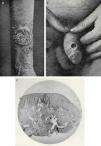
Regarding the book's discussion of acute and chronic pyoderma (in Chapters xvii and xviii, respectively), we will comment on the sections on “chronic vegetative papillomatous pyoderma with cystic epithelial reaction” and “chancriform pyoderma.” The first condition was described by de Azúa16 at the end of the 19th century in a series of patients with lesions similar to “vegetative epitheliomas” with a purulent exudate coming from hair follicles (Fig. 5A). The condition was quickly cured with antiseptic treatment and was initially called a pseudoinflammatory vegetative epithelioma. Histology showed a pseudoepitheliomatous hyperplasia with formation of keratotic cysts. However, these observations received little attention or acceptance abroad, as German authors claimed they had made the discovery first.
Chancriform pyoderma was described by Sánchez-Covisa and Bejarano6 in 2 boys with foreskin lesions resembling the chancres of primary syphilis. Later they found similar lesions in grown men (Fig. 5B), women, and girls and at multiple extragenital sites. In any case, attempts to find treponemes failed and serologies were negative. Another finding relevant to differential diagnosis was the absence of plasma cells in the infiltrate, which mainly consisted of polymorphonuclear cells (Fig. 5C). Staphylococcus bacteria were most often implicated, based on examination of lesion smears. Once again, a condition that was described first by Spanish authors would receive little international attention.
The last 2 chapters of the book deal with leprosy and tuberculosis.
Leprosy had been the subject of Sánchez-Covisa's talk on his induction into the Royal Academy of Medicine in Madrid on June 6, 1928.17 This disease was a genuine health problem in Spain (Fig. 6A), to the extent that the actual number of cases could be estimated to lie between 2000 and 2500, more than were suggested by official statistics (Fig. 6B). The authors of The Elements of Dermatology propose a systematic set of tests for diagnosing doubtful cases (Table 1).
Diagnostic Tests for Doubtful Cases of Leprosy, as Recommended by Sánchez-Covisa and Bejarano.
| 1. | Look for evidence of the bacillus in the nasal mucosa. |
| 2. | Examine the nasal mucosa after challenge with a tincture of iodine. |
| 3. | Examine suspicious lesions, obtaining punch biopsies with a capillary pipette. |
| 4. | Examine a smear from excised tissue. |
| 5. | Study tissues with acid-fast staining methods. |
| 6. | Study urine sediments, eventually distinguishing acid-fast bacilli grown in cultures inoculated with the material. |
| 7. | Study liquid aspirated from the testicles. |
| 8. | When necessary and if possible, biopsy and study the cubital nerve. |
They also provide a critical description of the various serology methods available, showing the results obtained in Spanish patients they treated or who were treated by other physicians, among them Drs Hombría, Enterría, González Medina, and Navarro Martín.
Great emphasis is placed on therapeutic hygiene and the careful organization of leprosy hospitals, given that the systemic treatments available at the time (arsenic, iodine, gold, antimony, timolol, or methylene blue) were not very effective.
Finally, they deal with cutaneous tuberculosis, which was also highly prevalent in Spain. The skin presentations of tuberculosis they describe are highly varied, classified as “1st, Acute tuberculous ulcer; 2nd, Fungoid tuberculosis; 3rd Verrucous tuberculosis; 4th, Tuberculosis luposa; 5th, Nodular tuberculosis; 6th, Exanthematous tuberculosis; and 7th, Abnormal forms of cutaneous tuberculosis.”
Curiously, the authors include erythematous tuberculosis under this last heading because it shared the geographic distribution of tuberculosis luposa, a preference for the female sex, the location of lesions, good response to treatment with gold, and the risk of consecutive epithelioma. They also mention finding many cases of tuberculosis luposa in association with other forms of tuberculosis.
Again the authors place great emphasis on hygiene and diet in the treatment of cutaneous tuberculosis, although they assert that the response is often unsatisfactory. Other treatments mentioned were gold salts, subcutaneous tuberculin, and local treatments such as Finsen light therapy, x-rays or radium treatments, electrocoagulation, dry ice, and pyrogallol. For small lesions they remark that “excision... combined with grafts and plasties... give surprisingly good therapeutic results in skilled hands.”
DiscussionTreatises on dermatology in Spanish prior to the appearance of The Elements of Dermatology fell into 3 broad categories:
- •
Mere translations of foreign texts. A noteworthy example in this group was Antonio Lavedán's 1798 translation of Plenck's Doctrina de Morbis Cutaneis.18
- •
True treatises on dermatology. In the middle of the 19th century, in the midst of the battle between the physiological doctrine of Alibert and the anatomical position of Willan, Spain saw the publication of 2 such works on skin diseases, one by Nicolás de Alfaro in 184019 and the other by Juan Luciano Murrieta in 1848.20 Both authors offered an interesting history of dermatology and analyzed the different nosological doctrines that reigned in their day.
- •
Lecture collections. Both Olavide in 186621 and Giné i Partagàs in 188022 published their dermatology lectures.
The Elements of Dermatology was a modern text, however. It was readable, well organized, and clearly sought to educate. Even though the target readers were students and general practitioners, the book delved deeply into the use of histology, pathology and the microbiologic methods of the day. The diseases were organized in keeping with the most advanced European currents in classification in the first third of the 20th century. The authors adopted Brocq's theory, based on both etiology and individual predisposition.
A major difference between this dermatology textbook and previous ones published in Spain was the large number of clinical and histologic photographs. Earlier book illustrations had been limited to reproductions of engravings and other figures. Sometimes these were impressive, such as those in Olavide's atlas of “clinical iconography” of skin diseases. Exceptionally, Giné i Partagàs's work on “clinical iconography” included 3 photographs at the end.22
The images in The Elements of Dermatology were so good that Gay Prieto23 would include some of them in his own book 6 years later. However, he very seldom cites Sánchez-Covisa or Bejarano directly in that work, merely mentioning them in connection with the vegetative lesions of bromism, the relation between psoriasis and joint disease, and potentially precancerous lesions. We may suppose that in the early years of the dictatorship it would have been difficult to obtain the authorities’ permission to publish a text that openly cited authors who had supported the Republic. As a result, the successors of Sánchez-Covisa and Bejarano consigned these men to oblivion: whether out of caution or ideology, they failed to cite their predecessors.
The lack of any chapters on venereology is very surprising. Although venereal diseases carried less weight in routine practice than they had in previous decades, they were still highly prevalent at this time. We will never know whether this area had been destined to become the subject of another manual.
Various explanations can account for the lack of new editions of the book. The authors’ exile—to Mexico and Venezuela—at a time when communication was not as easy as it is today would have made it hard for the co-authors to coordinate work on a second edition, and then Sánchez-Covisa died in 1944. In addition, a logistical difficulty for book publishers in the early postwar years was the severe shortage of paper and other printing supplies.
Aside from these problems, all those who expressed opposition to Franco's National Movement either through “concrete actions or serious passivity” were left, literally, “outside the law” according to the regime's Law of Political Responsibilities. The regime went further, zealously ferreting out and removing anything reminiscent of the Second Republic. Thus, in addition to the risk the authors would have run if they had returned and been brought before a military court, it is unlikely that the authorities serving the dictatorship would have allowed a new edition of the book to be published or sold.
Many volumes of the first edition of the textbook seem to have been destroyed along with many other books when the printer, Unión Poligráfica, was destroyed by howitzers in the last days of the Civil War, as reported by the press at the time.15,24 Thus, circulation was also severely restricted after existing copies were lost.
Concluding RemarksThe Elements de Dermatology was the first modern dermatology textbook written by Spanish authors and as such it should be remembered and given the recognition it deserves.
Its authors were progressive in assigning a prominent role to etiology and pathogenesis when they classified skin diseases, in drawing attention to advances in diagnostic methods and treatments, and in using photographs to teach dermatology.
However, Sánchez-Covisa, Bejarano, and their book were shunned as a result of the overwhelming social, cultural, and scientific upheaval brought about by the Civil War and the dictatorship it ushered into power.
Ethical DisclosuresProtection of human and animal subjectsThe authors state that no experiments were performed on humans or animals for this investigation.
Confidentiality of dataThe authors declare that they have followed the protocols of their hospitals concerning the publication of patient data and that all the patients included in this study were appropriately informed and gave their written informed consent.
Right to privacy and informed consentThe authors declare that no patient data appear in this article.
Conflicts of InterestThe authors declare that they have no conflicts of interest.
Please cite this article as: Leis-Dosil V, Garrido-Gutiérrez C, Díaz-Díaz R. Elementos de Dermatología. El legado de Covisa y Bejarano. Actas Dermosifiliogr. 2014;105:263–270.


