


Botulinum toxin A has emerged as an alternative treatment for patients who have contraindications to other therapies or as an adjuvant therapy in the treatment of androgenetic alopecia (AGA), although little is known about its safety and efficacy profile or the mechanism involved in this disease.
ObjectiveTo evaluate the efficacy profile of botulinum toxin as monotherapy for AGA and compare subcutaneous vs subcutaneous and intramuscular application.
MethodsWe conducted a 6-month, randomized, controlled trial to compare the efficacy of subcutaneous vs combined intramuscular and subcutaneous botulinum toxin application in men with AGA at a dermatology clinic in northeastern Mexico.
ResultsA total of 15 men, aged 27–63 years, diagnosed with androgenetic alopecia (AGA) classified as stages III to V on the Hamilton-Norwood scale, and with no prior treatment for, at least, 6 months prior to being included in the study, were randomly allocated following a computer-generated sequence created prior to the start of the study. The sequence was blinded to both patients and investigators. Participants received a single session of botulinum toxin, administered either subcutaneously and intramuscularly (Group A) or subcutaneously as monotherapy (Group B). Each patient received 100 units of botulinum toxin. When comparing the initial and follow-up trichoscopic photographs of the frontal and vertex regions 6 months after botulinum toxin application using Fotofinder Trichoscale®, no significant changes were observed in hair density or the vellus/terminal hair ratio in either Group A or Group B (p>0.05), and a significant reduction in hair thickness was detected in the frontal region of both groups. Terminal hair count in the frontal region decreased significantly in Group A (p=0.035), whereas the reduction in Group B was not statistically significant. In the occipital region, none of the evaluated parameters showed significant changes (p>0.05). Additionally, no significant differences were seen between the 2 treatment modalities 6 months after application (p>0.05).
ConclusionBased on our study, we did not find any supportive results for the use of botulinum toxin in the treatment of AGA.
La toxina botulínica tipo A ha surgido como un tratamiento alternativo para pacientes que tienen contraindicaciones para otras terapias o como un complemento en el tratamiento de la alopecia androgenética (AGA), aunque se sabe poco sobre su efectividad, seguridad y el mecanismo por el cual actúa en esta enfermedad.
ObjetivoEvaluar la eficacia de la toxina botulínica como monoterapia para la AGA y comparar la aplicación subcutánea frente a la aplicación subcutánea e intramuscular combinada.
MétodosSe llevó a cabo un ensayo clínico controlado, aleatorizado, de 6 meses de duración para comparar la efectividad de la aplicación de toxina botulínica subcutánea frente a la aplicación combinada intramuscular y subcutánea en pacientes masculinos con AGA en una clínica dermatológica del noreste de México.
ResultadosQuince pacientes masculinos, con edades entre 27 y 63 años, diagnosticados con alopecia androgenética (AGA) clasificada en los estadios III a V de la escala de Hamilton-Norwood, y sin tratamientos previos durante al menos seis meses antes de su inclusión, fueron asignados aleatoriamente utilizando una secuencia generada por computadora creada antes del inicio del estudio. La secuencia fue cegada tanto para los pacientes como para los investigadores. Los participantes recibieron una única sesión de toxina botulínica, administrada de forma subcutánea e intramuscular (Grupo A) o subcutánea como monoterapia (Grupo B). Cada paciente recibió un total de 100 unidades de toxina botulínica. Al comparar las fotografías tricoscópicas iniciales y de seguimiento de las regiones frontal y vértex, tomadas 6 meses después de la aplicación de toxina botulínica utilizando Fotofinder Trichoscale®, no se observaron cambios significativos en la densidad de cabello ni en la relación de vellos/terminales en ninguno de los grupos A o B (p>0.05). Sin embargo, se detectó una reducción significativa en el grosor del cabello en la región frontal en ambos grupos. Además, el conteo de pelos terminales en la región frontal disminuyó significativamente en el Grupo A (p=0.035), mientras que la reducción en el Grupo B no fue estadísticamente significativa. En la región occipital, ninguno de los parámetros evaluados mostró cambios significativos (p>0.05). Asimismo, no se encontraron diferencias significativas entre ambas modalidades de tratamiento 6 meses después de la aplicación (p>0.05).
ConclusiónEn este estudio no encontramos resultados que respalden el uso de toxina botulínica en el tratamiento de la AGA.

Androgenetic alopecia (AGA) is the most common type of hair loss in men.1 It has a higher incidence rate in Caucasians and is estimated to affect 50% and 73% of men older than 50 and 80 years, respectively.1,2 The pathogenesis of AGA is not fully understood; however, it is primarily mediated by dihydrotestosterone (DHT) which induces miniaturization of hair follicles, leading to progressive hair loss.1,2 DHT is produced by the action of 5α-reductase on testosterone in the cytoplasm of hair bulb cells and induces the production of transforming growth factor β1 (TGF-β1) in dermal papilla cells, suppressing the growth of follicular epithelial cells.3,4 Over time, this results in a progressive reduction in hair density, replacement of terminal hair with vellus hair, and irreversible damage to hair follicles, ultimately progressing to scarring alopecia.5
Recently, multiple effective treatments have been found for AGA, aiming to halt disease progression and, secondarily, to increase hair density.5,6 However, these treatments have limitations due to their adverse effects, duration of use, or invasiveness, prompting the search for new therapeutic options.3,6
Botulinum toxin A has emerged as an alternative treatment for patients who have contraindications to other therapies or as an adjuvant therapy in the treatment of AGA. This neurotoxin, produced by the bacterium Clostridium botulinum, selectively inhibits the presynaptic release of acetylcholine at the neuromuscular junction. It is widely used in dermatology to reduce the appearance of wrinkles, hyperhidrosis, scars, and temporomandibular disorders.7 Some studies have shown that it may be useful for treating AGA, although little is known about its safety and efficacy profile or the mechanism involved in this disease.
This study evaluated the efficacy profile of botulinum toxin as monotherapy for AGA and compared subcutaneous vs subcutaneous and intramuscular application. Hair density, hair thickness, and the ratio of vellus to terminal hairs were compared before and after treatment, as well as between the 2 application modalities.
MethodsStudy designWe conducted a 6-month, randomized, controlled study to compare the efficacy profile of subcutaneous vs combined intramuscular and subcutaneous botulinum toxin application in men with AGA at a dermatology clinic in northeastern Mexico. Patient recruitment spanned from January through February 2024. The study was approved by our hospital Ethics Committee. All participants gave their prior written informed consent.
Sample sizeThis pilot study aimed to assess the feasibility and potential effects of botulinum toxin A for treating AGA. A sample size of 7–8 participants per group was selected, appropriate for pilot studies to gather preliminary data and guide the design of a future, larger trial.
ParticipantsA total of 15 men aged 27–63 with AGA (Hamilton-Norwood scale III–V), who had not received any treatment for, at least, 6 months prior to inclusion, were randomized using a computer-generated sequence to receive a session of botulinum toxin either subcutaneously and intramuscularly (Group A) or subcutaneously as monotherapy (Group B). The sequence was blinded to both patients and investigators.
Exclusion criteria included the use of drugs or supplements that could influence hair metabolism, presence of infectious or inflammatory scalp conditions, any form of alopecia other than androgenetic alopecia (AGA), systemic conditions associated with telogen effluvium, or a documented history of hypersensitivity to botulinum toxin. Participants were allocated to treatment groups based on a predefined randomization sequence generated prior to starting the study.
Patients were required to attend the initial visit and 3 additional visits scheduled at 1, 3, and 6 months. Each visit included clinical and trichoscopic digital photographic follow-up using Fotofinder Trichoscale®.
Application of botulinum toxinTopical anesthesia with a cream containing 7% tetracaine and 23% lidocaine was applied 30min prior to the injection.
Botulinum toxin A (100U) was diluted in 1mL of 0.9% saline solution. Each patient received a total of 100 units administered with a 32G×4mm needle.
In Group A, consisting of 8 patients, 30 units of botulinum toxin were injected intramuscularly, bilaterally, into the frontal, temporal, and occipital muscles. Each muscle received 2 injections of 2.5 units each. Additionally, 70 units were administered subcutaneously in the frontal, parietal, and vertex regions of the scalp, with 30–40 injection points spaced 1.5–2cm apart, and 2 units injected at each point.
In Group B, 100 units were administered subcutaneously as described above: 2 units were injected at approximately 50 points spaced 1–1.5cm apart in the frontal, parietal, and vertex regions of the scalp.
Efficacy assessmentThe primary efficacy endpoint was any changes in hair density, hair diameter, and the ratio of vellus-to-terminal hairs in the frontal, midline, and vertex regions before and 6 months after the application of the botulinum toxin. A blinded clinical and trichoscopic analysis was performed using images from Fotofinder Trichoscale®.
The secondary efficacy endpoint was the global photographic assessment (GPA) conducted by 3 blinded dermatologists. They compared baseline photographs with those obtained at the final follow-up, 6 months after therapy. The assessment scale had 4 points: worsening (−1), stable (0), mild improvement (1), and marked improvement (2). An improvement of ≥1 grade on the GPA scale was defined as a marked improvement.
Additionally, patients assessed their improvement using a visual analog scale (0–10) by comparing their initial and final clinical photographs taken at the end of the 6-month follow-up.
Safety assessment included recording adverse events reported by patients and evaluated by the dermatologist during and after the treatment.
Statistical analysisAll statistical analyses were performed using SPSS (Statistical Package for the Social Sciences, version 23.0). Continuous variables are expressed as medians and interquartile ranges and categorical variables as whole numbers and percentages. Mann–Whitney U test and Fisher's exact test were used as hypothesis tests for continuous and categorical variables respectively. A p-value <0.05 was considered statistically significant.
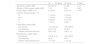
ResultsA total of 15 men, aged 27–63 participated in the study (8 patients were randomized to Group A to receive an intramuscular+subcutaneous dose of botulinum toxin in the scalp, and 7 to Group B to receive a subcutaneous dose). All patients completed the initial and follow-up visits. Demographic data and baseline characteristics were similar between the 2 treatment groups (Table 1). Efficacy results were evaluated 6 months after the botulinum toxin application (Table 2).
Demographics and baseline characteristics.
| IM+SC group | SC group | p-Value | |
|---|---|---|---|
| Age (years), median (IQR) | 42.5 (27.5) | 41 (28) | 0.77a |
| Duration of AGA (years), median (IQR) | 7 (4) | 5 (3) | 0.86a |
| Family history of AGA, n (%) | 8 (100%) | 4 (57.1%) | 0.077b |
| Hamilton–Norwood scale, n (%) | 1 | ||
| III | 1 (12.5%) | 1 (14.3%) | |
| III–V | 1 (12.5%) | 1 (14.3%) | |
| IV | 5 (62.5%) | 5 (71.4%) | |
| V | 1 (12.5) | 0 (0%) | |
| Frontal area, median (IQR) | |||
| Hair density | 173.3 (54.25) | 149.5 (68.6) | |
| Hair thickness | 40.5 (10.5) | 44 (21) | |
| Relation vellous/terminal hair | 0.84 (1.65) | 0.42 (1.11) | |
| Occipital area, median (IQR) | |||
| Hair density | 130.05 (41.5) | 142.8 (80.8) | |
| Hair thickness | 38 (6) | 38 (7) | |
| Relation vellous/terminal hair | 1.4 (1.64) | 1.3 (1.02) | |
IQR: interquartile range.
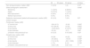
Perioperative pain assessment during, patient self-assessment, and clinical evaluation by dermatologist 6 months into therapy vs baseline.
| IM+SC group | SC group | p-Value | |
|---|---|---|---|
| Pain during procedure, median (IQR) | 1.5 (2.5) | 1 (3) | 0.72a |
| Global photographic assessment | 0.648b | ||
| Worsening | 1 (12.5%) | 2 (28.6%) | |
| Stable | 3 (37.5%) | 1 (14.3%) | |
| Mild improvement | 4 (50%) | 4 (57.1%) | |
| Marked improvement | 0 (0%) | 0 (0%) | |
| Subjective improvement (patient self-assessment), median (IQR) | 6.5 (3.5) | 7 (2.5) | 0.9a |
| Overall changes | |||
| Frontal area, median (IQR) | |||
| Δ Terminal hair | −26.6 (41.5) | −9 (34) | 0.082a |
| Δ Vellus hair | 10.5 (69.45) | 13 (54) | 0.862a |
| Δ Hair density | −16.05 (64.2) | 31 (87.5) | 0.452a |
| Δ Hair thickness | −5.5 (9) | −5 (7) | 0.86a |
| Δ Relation vellous/terminal hair | 0.6 (2.9) | 0.18 (0.68) | 0.42a |
| Occipital area, median (IQR) | |||
| Δ Terminal hair | 18 (26) | 14 (22) | 0.562a |
| Δ Vellus hair | −6.5 (30.5) | −15 (57) | 0.729a |
| Δ Hair density | 1.15 (14.35) | −021.1 (59.8) | 0.729a |
| Δ Hair thickness | 1 (7.5) | 0 (15) | 0.729a |
| Δ Relation vellous/terminal hair | −0.26 (1.26) | 0.021 (0.9) | 1a |
IQR: interquartile range.
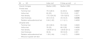
Comparison of trichoscopic images 6 months after botulinum toxin application in the frontal and vertex regions revealed varied results (Tables 2–4). While no significant changes were observed in hair density or the vellus/terminal hair ratio in either Group A or Group B (p>0.05), a significant reduction in hair thickness was detected in the frontal region of both groups. Additionally, terminal hair count in the frontal region decreased significantly in Group A (p=0.035), whereas the reduction in Group B was not statistically significant. In the occipital region, none of the evaluated parameters revealed any significant changes (p>0.05).
Baseline and 6-month follow-up changes in evaluated parameters in Group A (IM+SC).
| IM+SC | Index visit | Follow-up visit | p |
|---|---|---|---|
| Overall changes | Median (IQR) | Median (IQR) | |
| Frontal area | |||
| Terminal hair | 75.5 (48.5) | 44 (30.9) | 0.0357 |
| Vellus hair | 80 (55) | 79.5 (74.5) | 0.327 |
| Hair density | 173.3 (54.25) | 133.4 (52.6) | 0.5276 |
| Hair thickness | 40.5 (10.5) | 34.5 (5.5) | 0.0346 |
| Relation vellous/terminal hair | 0.84 (1.62) | 2.11 (2.1) | 0.4008 |
| Occipital area | |||
| Terminal hair | 49.5 (38.5) | 55.5 (32) | 0.2620 |
| Vellus hair | 78 (32) | 66.5 (35) | 0.4838 |
| Hair density | 130.05 (41.5) | 130.65 (51.5) | 0.5235 |
| Hair thickness | 38 (6) | 39.5 (9.5) | 0.831 |
| Relation vellous/terminal hair | 1.39 (1.64) | 1.14 (0.7) | 0.2626 |
| Wilcoxon signed rank test | |||
Bold values represent statistically significant differences (p < 0.05).
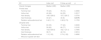
Baseline and 6-month follow-up changes in evaluated parameters in Group B (SC).
| SC | Index visit | Follow-up visit | p |
|---|---|---|---|
| Overall changes | Median (IQR) | Median (IQR) | |
| Frontal area | |||
| Terminal hair | 79 (24) | 78 (13) | 0.4990 |
| Vellus hair | 40 (69) | 64 (55) | 0.3517 |
| Hair density | 149.5 (68.6) | 157.2 (69.7) | 0.5754 |
| Hair thickness | 44 (21) | 40 (9) | 0.0176 |
| Relation vellous/terminal hair | 0.42 (1.11) | 0.98 (0.73) | 0.1763 |
| Occipital area | |||
| Terminal hair | 63 (35) | 77 (43) | 0.0630 |
| Vellus hair | 59 (62) | 66 (77) | 0.3105 |
| Hair density | 142.8 (80.8) | 162.7 (73.1) | 0.7995 |
| Hair thickness | 38 (7) | 42 (12) | 0.9314 |
| Relation vellous/terminal hair | 1.3 (1.02) | 0.74 (1.22) | 0.4990 |
| Wilcoxon signed rank test | |||
Bold values represent statistically significant differences (p < 0.05).
The median±interquartile range of hairs per square centimeter (hair density) in the frontal mid-region in Group A at the beginning of treatment and 6 months later was 173.3±54.25 and 133.4±52.6, respectively (p=0.527). Meanwhile, in Group B, it was 149.5±68.6 and 157.2±69.7 (p=0.575) at the start and after 6 months, respectively. In the occipital region, the hair density in Group A was 130.05±41.5 before treatment and 130.65±51.5 after treatment (p=0.523), while in Group B, it was 142.8±80.8 before treatment and 162.7±73.1 after treatment (p=0.7995).
The median±interquartile range of terminal hairs per square centimeter in the frontal mid-region in Group A at the start of the treatment and after 6 months was 75.5±48.5 and 44±30.9, respectively (p=0.035). Meanwhile, in Group B, it was 79±24 and 55.5±32 (p=0.262) at the beginning and 6 months later, respectively.
In the occipital region, the mean±standard deviation of terminal hairs per square centimeter was 49.5±38.5 and 55.5±32 (p=0.262) in Group A before and 6 months later, respectively. In Group B, it was 63±35 and 77±43 (p=0.063) at the beginning and 6 months later, respectively.
Hair thickness showed a statistically significant worsening in the frontal region over the 6-month period in the 2 groups. In Group A, it went from 40.5±10.5μm at baseline down to 34.5±5.5μm at the end of treatment (p=0.0346). Similarly, in Group B, it went from 44±21μm down to 40±9μm (p=0.0176). In the occipital region, no significant differences were observed before and after treatment in either Group A (p=0.831) or Group B (p=0.931).
The vellus/terminal hair ratio did not show any statistically significant differences before and after treatment in either Group A or Group B. In Group A, the median ratio in the frontal area was 0.84±1.62 at baseline and 2.11±2.16 months later (p=0.4), while in the occipital area, it was 1.39±1.64 at baseline and 1.14±0.7 after treatment (p=0.262). In Group B, the median ratio in the frontal area was 0.42±1.11 at baseline and 0.98±0.736 months later (p=0.176), while in the occipital area, it was 1.3±1.02 at baseline and 0.74±1.22 after treatment (p=0.499).
Additionally, no significant differences were found between the 2 groups in any of the evaluated parameters (p>0.05) (Table 2).
Global photographic assessmentThe global photographic assessment showed no statistically significant differences between the 2 groups (p=0.648) (Figs. 1 and 2). In the IM+SC group, 12.5% of patients showed worsening, 37.5% remained stable, and 50% showed mild improvement. In the SC group, 28.6% of patients showed worsening, 14.3% remained stable, and 57.1% showed mild improvement. There were no cases of marked improvement in either group (Table 2).
.")
.")
The median±interquartile range subjective improvement scores were 6.5±3.5 for the IM+SC group and 7±2.5 for the SC group, with no significant differences being reported between the 2 groups (p=0.9).
Safety and tolerabilityNo serious adverse events were reported in either group. Mild-to-moderate pain during the procedure was reported by 61.5% of patients. The mean pain scores during the procedure were 1.5±2.5 for the IM+SC group (Group A) and 1±3 for the SC (Group B), with no significant differences being reported between the 2 groups (p=0.72). No patient experienced headaches or itching associated with the application.
A post-hoc analysis resulted in a statistical power of 15.8% to detect a difference of global photographic assessment score.
DiscussionRecently, botulinum toxin A has emerged as a therapeutic option for patients with AGA. To this date, there are few studies supporting its efficacy profile, and although there are several hypotheses on the mechanism by which botulinum toxin may be beneficial in treating AGA, although it is not yet fully understood.
Former studies have suggested that patients with AGA have relative microvascular insufficiency and reduced blood flow in the most affected scalp regions, leading to a hypoxic state.8 This is partly due to the blood supply to areas affected by AGA being provided by the smaller branches of the internal carotid artery: the branches of the trochlear and supraorbital arteries.9 This hypoxic state is favored by the contraction of the frontal, occipital, periauricular, and temporal muscles.10 It has also been reported that the conversion of testosterone to DHT is favored by hypoxia.10,11
From this basis, the proposal arises that improving blood flow in the scalp can result in increased hair thickness and density, which would also increase the oxygen concentration, thus extending the anagen phase.9
Since scalp blood vessels reside in the intramuscular plane, botulinum toxin's muscle-relaxing effect could hypothetically boost blood flow and transcutaneous pO2 by easing pressure on musculocutaneous and perforating vessels.12
Additionally, increased blood flow might help wash away DHT resulting in increased hair thickness and density.12
It has also been reported that the subcutaneous administration of botulinum toxin A inhibits the secretion of TGF-β1 from dermal papilla cells, a pro-apoptotic molecule that suppresses the growth of follicular epithelial cells in AGA.2,9,13 Furthermore, TGF-β1 may be related to stiffness in areas affected by AGA, so botulinum toxin could contribute to an antifibrotic effect by decreasing this molecule.4
These are mechanisms of action of botulinum toxin found in studies on its application to the scalp in patients with AGA. To this date, very few studies on this topic have been published, and results on its efficacy profile are variable, with response rates reported up to 79.1% and an increase in hair count of 18%–20.9% at the end of patient follow-up.7
In former studies, intramuscular botulinum toxin was mainly applied to the frontal, temporal, periauricular, and occipital muscles, using a mean 50–150 units per session. The number of applications ranged from 1 to 6, with intervals of 4 weeks up to 3 months across sessions when multiple treatments were administered.10–12,14,15 Favorable outcomes were reported in 75%–79.1% of cases, with a mean increase in hair count of 10%–18%.3,12,14 One study evaluated the efficacy profile of intramuscular botulinum toxin using ultrasound and trichoscopy, reporting improvements in follicular width and length in the vertex region at 1 and 3 months post-treatment vs controls. However, no statistically significant differences were found in follicular or hair count.16
Similarly, Fernandes Melo et al. conducted a recent study comparing 50 units of botulinum toxin applied to one hemisphere of the scalp vs saline on the other. They found no significant benefits, or differences in terminal hair density, total hair density, vellus-to-terminal hair ratio, or cumulative hair thickness between hemispheres after 24 weeks. Additionally, a decrease in terminal hair density was observed in both the frontal and vertex scalp regions in both groups,17 which is similar to results from our study, where a decrease in terminal hairs was observed in frontal area in Group A.
Comparatively, the subcutaneous application of botulinum toxin for AGA has been evaluated in fewer studies. Tian et al. compared a total of 50 units of subcutaneous botulinum toxin to saline injections in patients concurrently receiving 1mg/day of finasteride and 5% topical minoxidil. Injections were administered to opposite hemispheres of the scalp. After 3 and 6 months, the treated hemisphere showed a significantly greater increase in hair density (29.31±11.94%) vs the control group (12.88±9.75%) (p<0.05).3
As far as we know, this is the first study ever conducted to compare intramuscular plus subcutaneous application (Group A) and subcutaneous application alone (Group B). At the 6-month follow-up, no statistically significant differences were observed in hair density or the vellus/terminal hair ratio from baseline to the end of treatment in either group, as analyzed using Trichoscale (p>0.05). However, a statistically significant decrease was found in hair diameter in the frontal region in both groups, and in the number of terminal hairs in Group A.
Our study found no greater effectiveness with the combined intramuscular and subcutaneous application of botulinum toxin vs subcutaneous application alone, although disease progression was observed in the frontal region in both Group A and Group B despite the intervention. Although various mechanisms of action have been proposed, neither administration route resulted in significant clinical or trichoscopic improvements. Therefore, our findings do not demonstrate a clear benefit from intramuscular, subcutaneous, or combined application as a treatment for AGA.
The global photographic assessment showed slight improvements in some patients, with 50% in Group A and 57.1% in Group B. Patients also reported a notable improvement on the visual analog scale; however, no statistically significant differences were observed across the groups. The discrepancy between objective and subjective results may be explained by a decrease in sebum and sweat production in treated patients. This cosmetic effect may give the impression of a slight increase in hair density, which could explain the discrepancy between subjective evaluation and trichoscopy. These results underscore the importance of integrating subjective evaluations with objective measurements to achieve more reliable and consistent results.
Other studies have reported additional benefits, such as less sebum production, scalp scaling, and pruritus after the application of botulinum toxin.3,10–14
In general, the application of botulinum toxin has proven to be a safe option.7,10 Former studies have reported no serious adverse effects, with only 1 patient developing a headache, mild dyspnea, and nausea, and the rest experiencing mild adverse effects, such as pain, erythema, or edema at the injection site.7 In our study, the only reported adverse effect was mild-to-moderate pain during application, with no serious adverse effects being reported.
This study has several limitations, including a heterogeneous patient population with a wide range of ages and varying degrees of AGA severity. Other limitations include the small number of participants typical of pilot studies, the 6-month follow-up, the absence of a placebo or control group, and the use of subjective outcome measures, such as patient self-assessments. Further studies with larger and more diverse patient populations are needed to improve the generalizability and external validity of these findings.
Although studies on the efficacy of botulinum toxin in AGA are highly heterogeneous regarding dosage, frequency, number, and location of injections, as well as study duration and evaluation parameters, its benefits appear to be minimal. Botulinum toxin has not been shown to significantly increase hair density, terminal hair count, or hair thickness.
Given its high cost and the limited therapeutic impact on AGA, alternative treatment options with greater demonstrated efficacy should be prioritized for these patients.
ConclusionsAlthough various mechanisms of action have been proposed, neither administration route resulted in significant clinical or trichoscopic improvements in our study. Consequently, our findings do not support a clear benefit derived from intramuscular, subcutaneous, or combined application as a treatment for AGA. Given its limited efficacy profile, dermatologists should carefully evaluate its cost–benefit ratio as an adjuvant therapy, particularly when alternative treatments with greater proven effectiveness remain available to us.
Conflict of interestThe authors declare that they have no conflict of interest.


