The main objective in dermatologic surgery is complete excision of the tumor whilst achieving the best possible functional and cosmetic outcome. In addition, we must take into account age, sex, and tumor size and site. We should also consider the patient's expectations, the preservation of the different cosmetic units, and the final cosmetic outcome.
The external nose is a facial esthetic unit that is divided into a number of cosmetic subunits that have a similar color, texture, and volume.
The ala nasi and the perialar region are anatomical areas that are commonly affected by neoplastic skin disease. This is a concave region at the confluence of the ala nasi, the lateral wall of the nose, the cheek, and the upper region of the lip. Its reconstruction is a challenge for the dermatologic surgeon, as poor planning of the technique can alter the alar sulcus, nasal symmetry, and the morphology of adjacent cosmetic units, and can produce functional changes such as alar retraction.1
The shark flap was first described by Cvancara and Wentzell2 in 2006. The flap was given this name because it is a myocutaneous island flap shaped like a shark. It is indicated for reconstruction of alar and perialar defects because, when it is performed, it involves a rotation that gives rise to the formation of an inverted cone that recreates the alar sulcus.
The classic flap is a myocutaneous flap that derives its blood supply from the levator labii superioris muscle and requires careful dissection to preserve the vascular pedicle. A variant with a subcutaneous pedicle with randomized vascularization has been described.3 This flap is simpler to perform and there is no increase in the risk of necrosis.
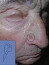
TechniqueWe present the case of an 84-year-old man with a basal cell carcinoma of 0.8×0.8cm on the right ala nasi (Fig. 1). Because of the site and size of the surgical defect created, we decided to use a shark flap for reconstruction.
The distance from the original alar sulcus to the medial border of the defect is measured and the flap is designed so that its superoanterior segment has the same width. The short limb of the flap is derived from the melolabial sulcus and the long limb will make use of the adjacent skin of the cheek. A flap with a subcutaneous pedicle is dissected, and the upper limb is then rotated through 90°, to lie perpendicular to the melolabial part of the flap and thus recreate the alar sulcus. The flap is then elevated and anchored in a superomedial direction. The rest of the flap is sutured, avoiding deep sutures that could increase the risk of necrosis.1
In our patient, the postoperative functional and cosmetic outcomes at 12 weeks were good (Fig. 2).
The surgical technique is shown in the video.
IndicationsThe shark flap is indicated for the repair of defects of the alar sulcus and perialar region. It has the advantage that it requires only a single operation, with no need for combination with other techniques (suture anchoring or grafts), and the functional and cosmetic results are good.2,4
Contraindications- -
This technique should not be used for large defects of the alar and perialar region as the cosmetic and functional outcomes will not be as good.
- -
This flap should not be used when cartilage is affected.3
- -
The main complication is flap necrosis, which can be prevented by performing correct dissection, achieving an appropriate length-to-width ratio of the flap, and by placing the sutures superficially to avoid damaging the blood supply (angular artery).
- -
Deformity of the ala nasi or of adjacent cosmetic structures through a poor design or execution of the flap.
- -
A trap door effect is possible because of the different thicknesses of the skin of the nose and cheek.2,4
The shark flap is a simple island flap that can be performed in a single operation. The flap recreates the alar sulcus with good cosmetic and functional results in comparison with other flaps employed in this region.
Conflicts of InterestThe authors declare that they have no conflicts of interest.
Please cite this article as: Pérez-Paredes MG, Valladares Narganes LM, Cucunubo HA, Rodríguez Prieto MÁ. Colgajo «en tiburón» para la reconstrucción de defectos de la región alar nasal. Actas Dermosifiliogr. 2014;105:709–711.


