


Anetoderma is a rare, benign disorder characterized microscopically by the loss of mid-dermal elastic fibers, resulting in well-circumscribed, skin-colored or gray-white atrophic macules or patches with a “sac-like” appearance.1 Anetoderma is classically divided into primary (idiopathic) and secondary anetoderma, with the former occurring in areas of previously normal skin and the latter developing at the site of a previous dermatosis.2 Secondary anetoderma has been associated with a broad spectrum of autoimmune, infectious, neoplastic and inflammatory dermatosis.1
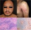
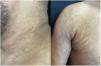
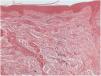
An otherwise healthy 35-year-old Indian man presented with a two-month history of multiple disseminated asymptomatic skin lesions. He had sex with women and reported multiple sex partners in the previous six months. No drug or medication use was elicited. He also denied any constitutional symptoms such as fever, malaise, or weight loss. On physical examination, there were multiple well-defined erythematous-violaceous, infiltrated, papules, plaques and nodules, symmetrically distributed on the face, neck, trunk and arms (Fig. 1A, B). Palmoplantar region and mucous membranes were not affected. Examination was otherwise unremarkable, without evidence of lymphadenopathy. Laboratory workup revealed a positive venereal disease research laboratory (VDRL) test result with a titer of 1:16 and a positive treponema pallidum hemagglutination (TPHA). Complete blood count and comprehensive metabolic panel were within reference range. Screening of other sexually transmitted diseases including human immunodeficiency virus (HIV), hepatitis C and hepatitis B virus was negative. Skin biopsy of an infiltrated erythematous plaque of the trunk was performed. Routine histopathological examination revealed a dense perivascular and periadnexal granulomatous infiltrate with plasma cells and histiocytes located in the superficial and deep dermis (Fig. 1C). Immunostaining using a polyclonal antibody for Treponema pallidum revealed the presence of multiple perivascular and intraepidermal spirochetes (Fig. 1D). Clinical, laboratory and histopathologic findings were thus compatible with the diagnosis of secondary syphilis. The patient was treated with a single dose of 2.4 million units of intramuscular benzathine penicillin G and clinical improvement was observed. However, six months later the patient presented to the clinic with asymptomatic, atrophic, wrinkled, skin-colored papules and plaques on the same locations that were previously affected by the syphilitic rash (Fig. 2). VDRL and viral serologies were negative. Skin biopsy was performed and histopathological examination with elastic tissue staining revealed a significant reduction of elastic fibers and a mild superficial perivascular lymphocytic infiltrate (Fig. 3). As such, clinical and histopathological findings confirmed the diagnosis of anetoderma due to secondary syphilis.
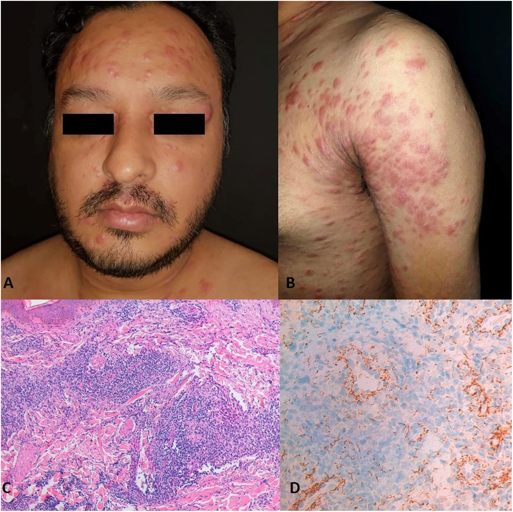
Multiple erythematous-violaceous, infiltrated, papules, plaques and nodules, symmetrically distributed on the face, neck, trunk and arms (A, B). Hematoxylin & eosin stain (100×) shows a dense perivascular and periadnexal granulomatous infiltrate with plasma cells and histiocytes in the superficial and deep dermis (C); The T. pallidum immunostain (100×) robustly highlights perivascular and intraepidermal spirochetes (D).


Clinical manifestations of secondary syphilis can be varied and often represent a diagnostic challenge. The most commonly observed clinical presentation (80%) is a generalized, non-pruritic papulosquamous eruption that typically affects palms and soles, accompanied by flu-like symptoms and generalized lymphadenopathy.3 However, a broad spectrum of cutaneous manifestations has been described in literature, frequently leading to misdiagnosis and, for that reason, secondary syphilis has been known as the “great imitator”. Notably, papulonodular presentation of secondary syphilis is uncommon, with no more than 30 reported cases.3 In our patient, differential diagnosis included cutaneous lymphoma, subacute cutaneous lupus erythematous, Sweet syndrome and cutaneous sarcoidosis. Those were excluded by clinical history, laboratory investigations and routine histopathology. At the time of diagnosis of syphilis, there was no other dermatologic process that could be the cause of anetoderma.
Usually, after appropriate treatment with penicillin, syphilitic lesions resolve without scarring.4 Secondary anetoderma due to syphilis has been poorly mentioned in literature. It may present in all stages of the disease, although it seems to be more common on the secondary stage and in HIV-infected patients.4 Even though there are multiple diseases that can evolve to anetoderma, its pathophysiology is still not fully understood. The decrease in elastic tissue may be due to either defective elastin synthesis, uncontrolled production of elastolytic enzymes, loss of elastolytic enzyme inhibitors, elastophagocytosis or degeneration of elastic fibers secondary to local ischemia induced by microthrombosis in dermal vessels.5 In addition, demonstration of fragmented elastic fibers present within macrophages by electron microscopic examination suggests that granulomatous diseases could evolve to anetoderma.2 Our case supports this hypothesis, since it presented with a granulomatous pattern in histopathological examination of syphilitic skin lesions.
There is no satisfactory treatment available for established anetoderma lesions.5 Cryotherapy, intralesional steroids and resurfacing laser have been used with variable success.5 Since the problem was solely cosmetic in nature, our patient declined any treatment attempt.
In conclusion, the present case highlights the occurrence of anetoderma after adequately treated papulonodular secondary syphilis and should further alert dermatologists for this rare potential association.
FundingNo funding has been received for the completion of this article.
Conflict of interestsThe authors declare that they have no conflict of interest.


