

Melanoma incidence continues to rise in different populations and ethnic groups worldwide.1–3 Epidemiological data and known risk factors for melanoma are mostly based on studies of US, Australian, and European populations. Few population-based studies have been conducted in Latin American or Hispanic communities.4
Latin America is known for its significant ethnic diversity secondary to interracial relationships that vary from one country to the next depending on population structure and migration history. In Colombia, for example, the population results from interactions between indigenous/native populations, Spanish people, and Africans, with mestizos representing the largest segment. The aim of this study was to identify possible risk factors for cutaneous melanoma in a Colombian population in the city of Medellín, Colombia.
We conducted a retrospective age- and sex-matched case-control study. Cases were patients with a histopathologically confirmed diagnosis of in situ or invasive melanoma, while controls were randomly selected patients without a personal history of melanoma who were seen for any dermatologic condition. The ratio of cases to controls was 1:2. We analyzed the medical records of patients older than 18 years seen at Clínica Aurora, a specialized skin cancer center, in Medellín between May 2014 and October 2017. We included both incident and prevalent cases of melanoma. In other words, we studied patients newly diagnosed with melanoma during the study period and those with an existing diagnosis. The required sample size was estimated at 62 cases and 125 controls using an alpha error of 5%, a beta error of 20% (95% confidence level and 80% statistical power) and an odds ratio (OR) of 3 associated with the presence of multiple melanocytic nevi as the main risk factor for melanoma. The calculations were performed in the statistical software program Epi Info.
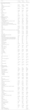
We analyzed the medical records of 187 patients (62 cases and 125 controls). Their phenotypic, sociodemographic, and sun exposure characteristics are given in Table 1. Table 2 summarizes the characteristics of the patients with melanoma. Table 3 presents the results of the bivariate and multivariate analyses.
Sociodemographic, Phenotypic, and UV Radiation Exposure Characteristics.
| Variable | Controls | Cases | Total |
|---|---|---|---|
| N = 125 (%) | N = 62 (%) | N = 187 (%) | |
| Sociodemographic characteristics | |||
| Sex | |||
| Male | 61 (49) | 35 (56) | 96 (51) |
| Female | 64 (51) | 27 (44) | 91 (49) |
| Median age (interquartile range), ya | 53 (37-63) | 53 (35-66) | |
| Civil status | |||
| Married | 93 (74) | 44 (71) | 137 (73) |
| Single | 32 (26) | 18 (29) | 50 (27) |
| Place of residence | |||
| Urban | 106 (85) | 58 (94) | 164 (88) |
| Rural | 15 (12) | 2 (3) | 17 (9) |
| Abroad | 4 (3) | 2 (3) | 6 (3) |
| Social security regime | |||
| None | 10 (8) | 7 (11) | 17 (9) |
| Subsidized | 1 (1) | 1 (2) | 2 (1) |
| Contributive | 19 (15) | 7(11) | 26 (14) |
| Private | 95 (76) | 47(76) | 142 (76) |
| Phenotypic characteristics | |||
| Red/blonde hair | 13 (10) | 7 (11) | 20 (10) |
| Blue/green/gray eyes | 22 (18) | 13 (21) | 35 (19) |
| Facial freckles | 4 (3) | 0 (0) | 4 (2) |
| Fitzpatrick skin type I or II | 34 (27) | 10 (6) | 44 (23) |
| > 5 clinically dysplastic nevi or at least 2 per condition | 15 (12) | 18 (29) | 33 (17) |
| Sun-damaged skin | 103 (82) | 46 (74) | 149 (80) |
| Personal history of melanoma | 5 (4) | 5 (8) | 10 (5) |
| Personal history of nonmelanoma skin cancer | 23 (18) | 6 (10) | 29 (15) |
| Family history of melanoma in a first- to third-degree relative | 14 (11) | 10 (16) | 24 (13) |
| Number of common nevi | |||
| 0-20 | 44 (35) | 8 (13) | 52 (28) |
| 20-50 | 32 (25) | 10 (16) | 42 (22) |
| 50-100 | 17 (14) | 12 (19) | 29 (16) |
| > 100 | 27 (22) | 11 (18) | 38 (20) |
| Not described | 5 (4) | 21 (34) | 26 (14) |
| Number of nevi on trunk and upper extremities (based on photographs) | |||
| 0-20 | 45 (36) | 8 (13) | 53 (29) |
| 20-50 | 32 (26) | 12 (19) | 44 (23) |
| 50-100 | 23 (18) | 11 (18) | 34 (18) |
| > 100 | 18 (14) | 10 (16) | 28 (15) |
| Not described | 7 (6) | 21 (34) | 28 (15) |
| Nevi: predominant dermoscopic pattern | |||
| Reticular diffuse | 78 (62) | 24 (38) | 102 (54) |
| Reticular patchy | 4 (3) | 0 (0) | 4 (2) |
| Peripheral reticular pattern with central hypopigmentation | 0 (0) | 0 (0) | 0 (0) |
| Peripheral reticular pattern with central hyperpigmentation | 0 (0) | 1 (2) | 1 (1) |
| Peripheral reticular pattern with central hypopigmentation | 0(0) | 0 (0) | 0 (0) |
| Homogenous pattern | 2 (2) | 0 (0) | 2 (1) |
| Peripheral globules | 1 (1) | 0 (0) | 1 (1) |
| Globular | 1 (1) | 0 (0) | 1 (1) |
| Two components | 7 (6) | 5 (8) | 12 (6) |
| Multiple components | 0 (0) | 1 (2) | 1 (1) |
| Not described | 32 (26) | 31 (50) | 63 (33) |
| Congenital melanocytic nevus | |||
| None | 117 (93) | 59 (96) | 176 (94) |
| Small (< 1.5 cm) | 8 (6) | 0 (0) | 8 (4) |
| Median (1.6-19.9 cm) | 0 (0) | 2 (3) | 2 (1) |
| Large (> 20 cm) | 0 (0) | 1 (1) | 1 (1) |
| Solar lentigines | |||
| In 1 area | 66 (53) | 32 (52) | 98 (52) |
| In 2 areas | 44 (35) | 21 (34) | 65 (35) |
| In ≥ 3 areas | 15 (12) | 8 (13) | 23 (12) |
| Not described | 0 (0) | 1 (1) | 1 (1) |
| Actinic keratosis | |||
| None | 104 (83) | 47 (76) | 151 (81) |
| < 10 | 17 (14) | 7 (11) | 24 (13) |
| > 10 | 4 (3) | 8 (13) | 12 (6) |
| History of another cancer | |||
| No | 119 (95) | 61 (99) | 180 (96) |
| Yes | 6 (5) | 1 (1) | 7 (4) |
| Chronic immunosuppression | |||
| No | 125 (100) | 62 (100) | 187 (100) |
| Yes | 0 (0) | 0 (0) | 0 (0) |
| Genodermatosis | |||
| No | 125 (100) | 62 (100) | 187 (100) |
| Yes | 0(0) | 0 (0) | 0 (0) |
| Parkinson disease | |||
| No | 124 (99) | 62 (100) | 186 (99) |
| Yes | 1 (1) | 0 (0) | 1 (1) |
| UV radiation exposure | |||
| Skin reaction to sunlight | |||
| No effect | 0 (0) | 0 (0) | 0 (0) |
| Tan | 17 (14) | 12 (19) | 29 (16) |
| Mild sunburn then tan | 75 (60) | 30 (48) | 105 (56) |
| Sunburn without blisters | 29 (23) | 13 (21) | 42 (22) |
| Sunburn with blisters | 3 (2) | 1 (2) | 4 (2) |
| Not described | 1 (1) | 6 (10) | 7 (4) |
| Lifetime history of sunburn | |||
| No episodes | 68 (54) | 13 (21) | 81 (43) |
| < 3 episodes | 34 (28) | 25 (40) | 59 (32) |
| > 3 episodes | 22 (17) | 6 (10) | 28 (15) |
| 1 (1) | 18 (29) | 19 (10) | |
| Sunburn before age of 20 years | |||
| No | 74 (59) | 16 (26) | 90 (48) |
| Yes | 50 (40) | 29 (47) | 79 (42) |
| Not described | 1 (1) | 17 (27) | 18 (10) |
| Frequency of sunscreen use | |||
| Never | 17 (13) | 7 (11) | 24 (13) |
| Only during intense sun exposure | 14 (11) | 3 (5) | 17 (9) |
| Occasional | 22 (18) | 11 (18) | 33 (17) |
| Once a week | 1 (1) | 0 (0) | 1 (1) |
| Once a day | 66 (53) | 29 (47) | 95 (51) |
| At least twice a day | 5 (4) | 0 (0) | 5 (3) |
| Not described | 0 (0) | 12 (19) | 12 (6) |
| Frequency of use of other sun protection measures | |||
| Never | 13 (11) | 8 (13) | 21 (12) |
| Sometimes | 53 (42) | 25 (40) | 78 (42) |
| Often | 56 (45) | 13 (21) | 69 (37) |
| Always | 3 (2) | 0 (0) | 0 (0) |
| Not described | 0 (0) | 16 (26) | 16 (9) |
| Lifetime use of physical sun protection measures | |||
| 1 method | 24 (19) | 19 (31) | 43 (23) |
| 2 methods | 89 (71) | 27 (43) | 116 (62) |
| 3 or more methods | 12 (10) | 0 (0) | 12 (6) |
| Not described | 0 (0) | 16 (26) | 16 (9) |
| Use of sunscreen before age of 18 years | |||
| Never | 47 (38) | 20 (32) | 67 (36) |
| Sometimes | 65 (52) | 24 (39) | 89 (47) |
| Often | 1 (1) | 0 (0) | 1 (1) |
| Always | 0 (0) | 0 (0) | 0 (0) |
| Not described | 12 (9) | 18 (29) | 30 (16) |
| Use of tanning booths | |||
| Never | 111 (89) | 39 (63) | 150 (80) |
| Yes, before age of 25 years | 6 (5) | 3 (5) | 9 (5) |
| Yes, after age of 25 years | 8 (6) | 4 (6) | 12 (6) |
| Not described | 0 | 16 (26) | 16 (9) |
| History of sunburn | |||
| None | 84 (67) | 17 (28) | 101 (54) |
| Intermittent | 31 (25) | 31 (50) | 62 (33) |
| Chronic | 9 (7) | 2 (3) | 11 (6) |
| Not described | 1 (1) | 12 (19) | 13 (7) |
| At least 1 modifiable factorb | 101 (54) | ||
| At least 1 nonmodifiable factorb | 48 (26) | ||
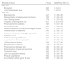
Characteristics of Patients With Melanoma (n = 62).
| Variable | N = 62 (%) |
|---|---|
| Person who detected the melanoma | |
| Patient | 5 (8) |
| Dermatologist | 56 (90) |
| Other physician | 1 (2) |
| Tumor site | |
| Extremities | 24 (39) |
| Trunk | 21 (34) |
| Head and neck | 13 (21) |
| Abdomen | 4 (6) |
| Dermoscopic features | |
| Atypical network | 22 (35) |
| Atypical globules | 15 (24) |
| Atypical vessels | 9 (15) |
| Regression | 7 (11) |
| Crystalline structures | 6 (10) |
| Multicomponent | 31 (50) |
| Not described | 13 (21) |
| Histologic features | |
| Melanoma in situ | 41 (66) |
| Invasive | 21 (34) |
| Breslow thickness < 0.8 mm | 15 (24) |
| Breslow thickness > 0.8 mm | 6 (10) |
| Ulceration | 1 (2) |
| Regression | 5 (8) |
| Perineural invasion | 1 (2) |
| Lymphovascular invasion | 0 |
| Microsatellites | 0 |
| Histologic subtype | |
| Superficial spreading | 21 (34) |
| Nodular | 6 (10) |
| Lentigo maligna | 8 (13) |
| Acral lentiginous | 2 (3) |
| Other: blue nevus–like | 1 (2) |
| Mitoses/mm2 | |
| Not applicable | 39 (63) |
| < 1 | 11 (18) |
| > 1 | 12 (19) |
| Growth phase | |
| Radial | 53 (85) |
| Vertical | 9 (15) |
| Staging | |
| In situ | 41 (66) |
| I | 16 (26) |
| II | 2 (3) |
| III | 2 (3) |
| IV | 1 (2) |
Bivariate and Multivariate Analysis of Risk Factors for Melanoma.
| Bivariate analysis | P Value | Odds Ratio (95% CI) |
|---|---|---|
| Hair color | ||
| Blonde/red | .802 | 1 (0.4-3.1) |
| Light Fitzpatrick skin type | .279 | 1 (0.5-2.5) |
| Eye color | ||
| Blue/green/gray | .547 | 1.3 (0.6-2.8) |
| Personal history of squamous cell carcinoma | .733 | 1.3 (0.4-5.0) |
| Use of tanning booths | .779 | 1.4 (0.3-5.9) |
| Frequency of tanning booth use | .567 | 1.42 (0.5-4.9) |
| Presence of actinic keratosis | .242 | 1.5 (0.7-3.3) |
| No. of common nevi | .085 | 1.7 (0.6-4.8) |
| History of melanoma in a first- to third-degree relative | .767 | 1.7 (0.5-6.1) |
| Personal history of melanoma | .245 | 2.1 (0.6-7.5) |
| Number of nevi on trunk and upper extremities | .158 | 2.1 (0.8-5.7) |
| Dysplastic nevus | .022 | 2.5 (1.1-5.7) |
| Sunburn before age of 20 years | .005 | 2.6 (1.3-5.4) |
| ≥ 1 predominant dermoscopic pattern | .06 | 2.9 (0.9-9.6) |
| Lifetime history of sunburn | .003 | 3.8 (1.7-8.4) |
| Recreational sun exposure | < .001 | 4.9 (2.4-10.1) |
| Presence of sun-damaged skin | .246 | 0.6 (0.3-1.3) |
| Personal history of basal cell carcinoma | .037 | 0.2 (0.0-0.9) |
| Congenital nevus | .502 | 0.4 (0.1-2.4) |
| Use of sun protection methods | .978 | 0.9 (0.4-1.9) |
| Physical sun protection | .040 | 0.3 (0.2-0.8) |
| Sun protection before age of 18 years | .660 | 1.1 (0.6-2.4) |
| Multivariate analysisa | OR (95% CI) |
|---|---|
| History of sunburn | 5.63 (2.2-14.6) |
| History of reactive sun exposure | 4.17 (1.8-9.6) |
| Use of 2 or more physical sun protection measures | 0.19 (0.1-0.5) |
χ2 test or Fisher exact test for counts below 5. Logistic regression was used for polytomous variables. Variables with a P value < .25 (Hosmer-Lemeshow goodness of fit test) were entered into the multivariate analysis.
Similarly to previous reports in the literature,5–7 our multivariate analysis showed that patients with melanoma were more likely to have a history of recreational or intermittent sun exposure (OR = 4.2; 95% CI, 1.8-9.6) and a lifetime history of sunburn (OR = 5.6; 95% CI, 2.2-14.6), suggesting that these patterns of exposure are risk factors for melanoma in our study population. In contrast, the use of 2 or more sun protection measures exerted a protective effect (OR = 0.2; 95% CI, 0.1-0.5). Unlike other studies, we did not observe an association between number of common or atypical nevi and melanoma risk.5,8 We observed a protective effect for a personal history of basal cell carcinoma, possibly because the control population was selected from a specialized center where patients with skin cancer predominate and basal cell carcinoma would be expected to be relatively common.
Our study has some limitations. First, the controls were selected from a referral center for patients with skin cancer and their exposure habits may not be representative of the general population in the area, possibly resulting in an overestimation of the proportion of patients with a history of skin cancer and creating a bias in our results for use of sun protection measures, access to health care, and level of education. Second, recall bias may have affected the accuracy or completeness of important data for our analyses, as some of the information was based on subjective perceptions of past events.
In conclusion, recreational or intermittent sun exposure and a lifetime history of sunburn were risk factors for melanoma in our study population, while use of 2 or more sun protection measures exerted a protective effect. Our findings are consistent with reports from different regions in the world, indicating that UV radiation plays an important role in populations other than Whites and that primary prevention efforts are also essential for preventing skin cancer in mestizo populations such as those in Colombia.
Conflicts of InterestThe authors declare that they have no conflicts of interest.
The following is Supplementary data to this article:

Please cite this article as: Aguirre LM, Muñoz AM, Aluma-Tenorio MS, Jaimes N. Factores de riesgo para melanoma en una población latinoamericana: estudio de casos y controles. Actas Dermosifiliogr. 2021;112:943–949.


