


Urethritis is a common reason for consultation in Venereology. Early diagnostic orientation facilitates the rational use of antibiotics. Although nucleic acid amplification techniques have accelerated microbiological diagnosis, their results are not immediate, and therefore direct visualization techniques still play a clear role in daily clinical practice. In this context, direct microscopic examination of urethral samples stained with methylene blue is a valuable, simple tool that provides virtually immediate results.
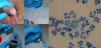
The procedure consists of obtaining a sample of urethral exudate with a swab (Fig. 1A) and spreading it onto a glass slide (Fig. 1B). After allowing it to air-dry for 1–2min, the slide is immersed in a methylene blue solution (Fig. 1C) and then rinsed with running water (Fig. 1D). Once the excess water has been removed, direct microscopic visualization using immersion oil is performed. In cases of gonococcal infection, this will reveal the presence of numerous polymorphonuclear leukocytes and intracellular diplococci (Fig. 1E).

We believe this method is of interest to dermatovenereologists because it is easy to implement and, in most cases, provides adequate results for guiding empirical treatment.


