We report the case of a 62-year-old Caucasian female, with an 8-year history of Waldenström’s macroglobulinemia (WM), under treatment with cyclophosphamide and prednisone, and a 1-year history of stage IV colon adenocarcinoma. The patient was referred to our Dermatology Department due to the recent occurrence of ill-defined, erythematous, infiltrated patches on the extensor surface of both arms, painful upon palpation (Fig. 1). She had no history of previous dermatological disorders and no triggering factors were identified. Cachexia and hepatosplenomegaly were also apparent, but the rest of the physical examination was unremarkable. Laboratory results showed a normocytic, normochromic anemia (hemoglobin 9.1 × 10 g/L), hypoalbuminemia (25.8 g/L), high levels of serum immunoglobulin M (IgM; 70.50 g/L) and of several tumor markers, namely cancer antigen (CA) 125, CA 19−9, CA 72−4 and carcinoembryonic antigen (CEA).
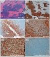
As the clinical findings were non-specific, we performed a deep skin biopsy of one lesion. The histopathological examination showed a diffuse, dense dermal and subcutaneous infiltration of small lymphocytes, lymphoplasmacytoid cells and plasma cells, staining positive for CD20, CD79a, CD138 and IgM, with kappa light chain restriction (Fig. 2), and negative for several T-cell markers, namely CD3, CD4 and CD8. These findings confirmed the diagnosis of specific skin infiltration by WM (lymphoplasmacytic lymphoma). Another scheme of systemic therapy directed to WM (bortezomib and dexamethasone) was started, together with topical clobetasol propionate on the skin lesions for local symptomatic relief, without significant clinical improvement, however. The progression of two concurrent advanced oncological diseases – colon cancer and WM – led to a rapidly progressive clinical deterioration that culminated in the patient’s death few months later.
Histopathological findings in skin lesion biopsy: Presence of a diffuse, dense dermal and subcutaneous infiltration (a) by small lymphocytes, lymphoplasmacytoid cells, and plasma cells, staining positive for CD20 (b), CD138 (c) and IgM (d), with kappa light chain restriction (e, f), confirming neoplastic cutaneous involvement by Waldenström’s macroglobulinemia.
WM is a lymphoproliferative disease, characterized by clonal proliferation of lymphoplasmacytoid cells producing a monoclonal IgM protein.1–3 A broad spectrum of cutaneous disorders has been associated with this monoclonal gammopathy, including non-neoplastic and neoplastic skin manifestations,1–3 which occur in only approximately 5% of the patients with WM.3 These can be due to several pathogenic mechanisms, namely: 1) specific cutaneous infiltration by neoplastic cells or deposition of their cellular products, particularly monoclonal IgM (“macroglobulinemia cutis”); 2) secondary to paraproteinemia, including mucocutaneous manifestations of hyperviscosity, cryoglobulinemia or autoimmune phenomena; 3) miscellaneous manifestations of uncertain etiology, as urticaria and amyloid light-chain (AL) amyloidosis.1–3
Specific infiltration of the skin by proliferating lymphoplasmacytoid neoplastic cells is, in fact, the rarest cutaneous manifestation of WM, with only few similar cases having been reported to date, but it should be considered in the differential diagnosis of asymptomatic or symptomatic, infiltrated skin lesions in these patients.1–6 Histopathological examination of skin specimens with immunoperoxidase stains is key in establishing the correct diagnosis.2 These lesions result from skin infiltration by malignant cells, and treatment should therefore target the underlying hematological malignancy,1 although clinical responses have been inconsistent.2 Interestingly, the presence of cutaneous involvement does not appear to be clearly related with disease course and progression, according to the available literature.2 Nevertheless, further research is warranted.
We report this clinical case to draw attention to an uncommon dermatological expression of a systemic hematologic malignancy and the importance of considering such possibility in appropriate clinical settings.
Please cite this article as: Valejo Coelho M.M., João A., Rocha Páris F. Infiltración neoplásica cutánea por un linfoma linfoplasmocítico en una paciente con macroglobulinemia de Waldenström. Actas Dermosifiliogr. 2020;111:528–531.


