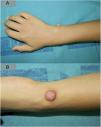
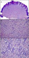
An 8-year-old boy with no medical history of interest was seen for an asymptomatic, fast-growing lesion on the right wrist that had appeared 3 months earlier. The lesion consisted of a firm, pink nodule (15 × 8 mm) with a slightly hyperkeratotic surface (Fig. 1). Histology showed a nodular tumor comprised of large nests of epithelioid and spindle cells that extended as far as the deep dermis. Neither necrosis nor mitotic figures were observed, and a diagnosis of Spitz nevus (SN) was established (Fig. 2).
SN is a consequence of melanocyte proliferation and is generally acquired, although congenital cases have been described.1 It typically manifests as soft, pinkish or homogeneously pigmented papules or nodules of 5–10 mm with clearly defined margins. Commonly affected areas include the lower extremities and the head and neck.2
Large SN with a verrucous appearance is infrequent. In such cases other diagnoses (common wart, molluscum contagiosum, etc.) should be considered. Histopathology is essential in cases of progressively growing lesions with an inconclusive clinical diagnosis.
Please cite this article as: Conde-Ferreirós A, Velasco-Tirado V, Santos-Briz Á, Yuste-Chaves M. Nevus de Spitz verrucoso en muñeca derecha. Actas Dermosifiliogr. 2021;112:266–267.


