
Milia are benign epidermal cysts that can present as an isolated finding or associated with other clinical alterations. When found in groups on an erythematous base, the lesions are called milia en plaque.
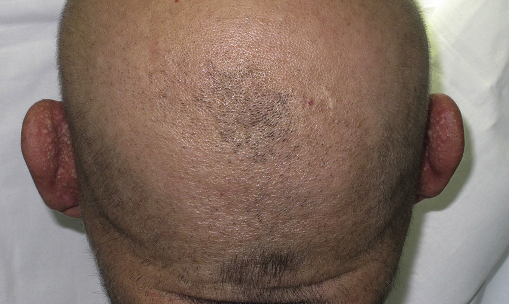
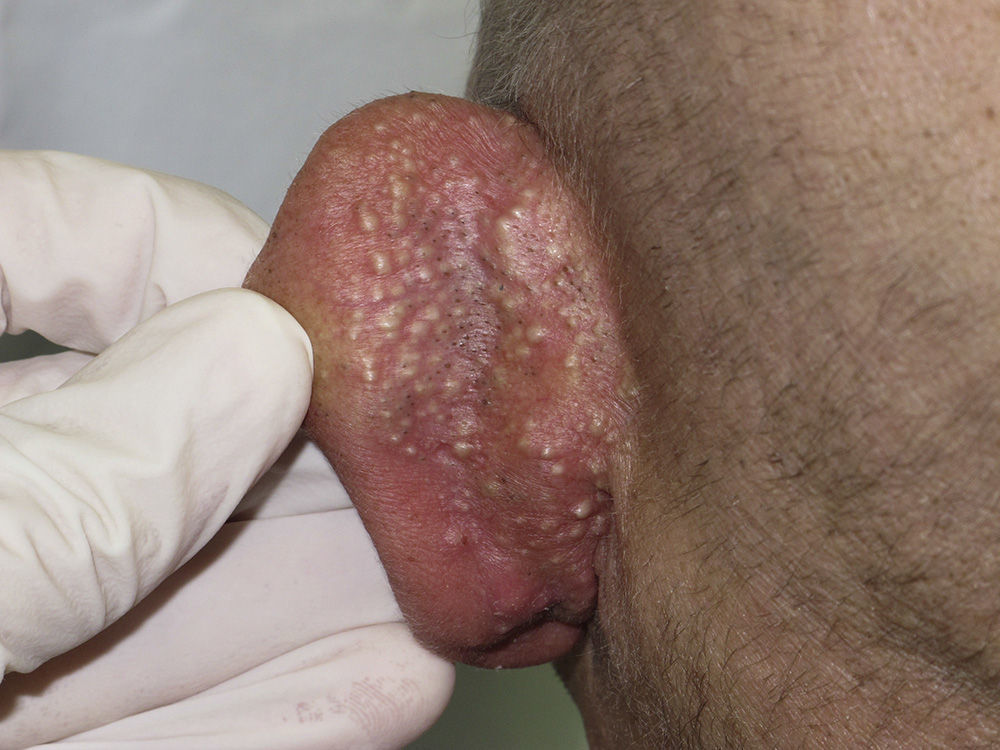
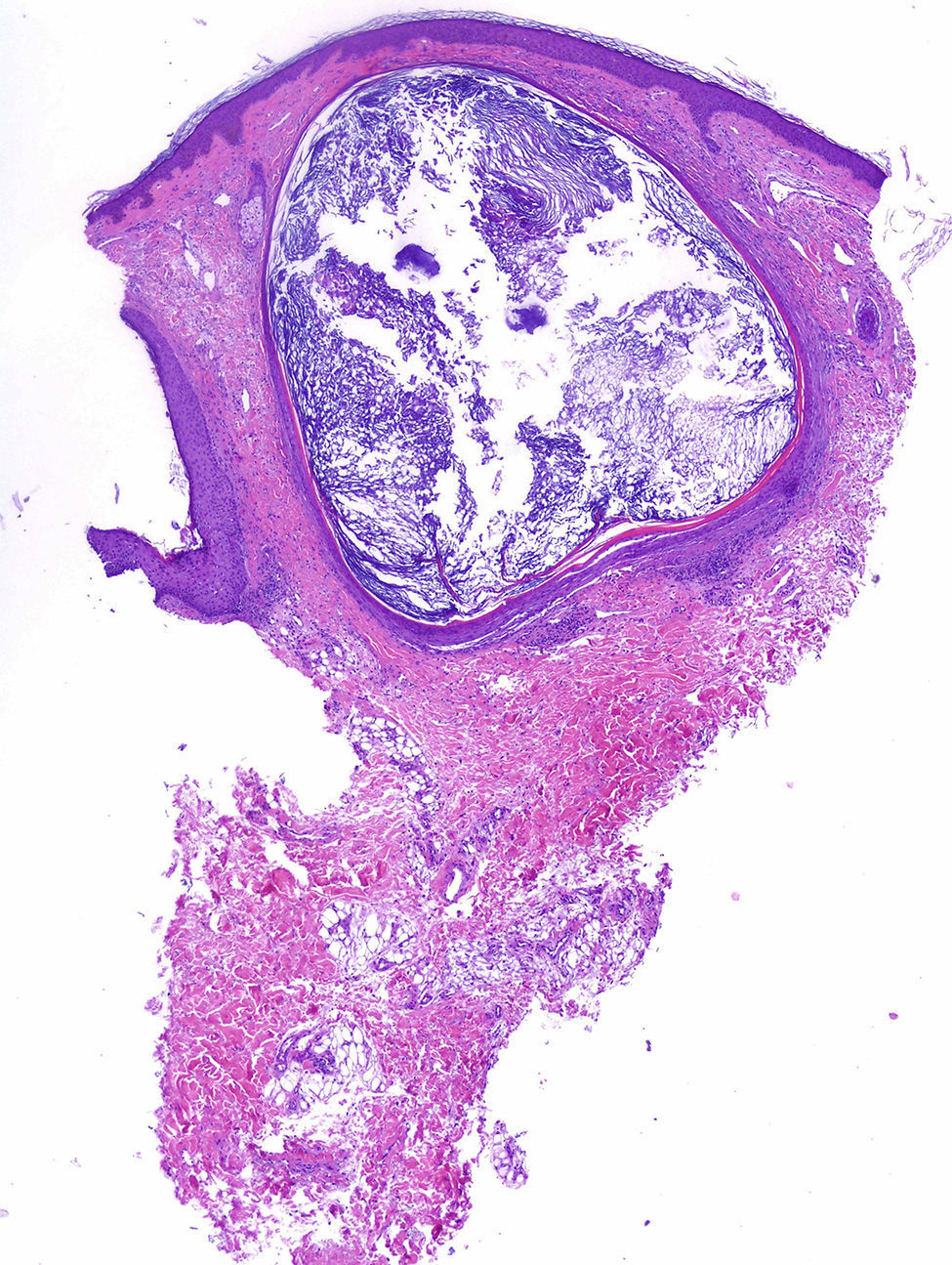
We present the case of a 48-year-old man with history of adenocarcinoma of the lung with cerebral metastases for which he received palliative treatment with holocranial radiotherapy at a dose of 30Gy in 10 fractions. The radiation fields included the auricles of the ear (2 opposing lateral photon beams to the central nervous system); the total dose received by the auricles of the ear was calculated as between 20 and 25Gy. The patient came to our outpatient clinic for asymptomatic lesions that had arisen on the posterior aspect of the auricles of both ears 3 months earlier. Since his youth he had occasionally presented isolated lesions of a similar appearance, but the multiple lesions had developed a month after the radiotherapy. On examination, multiple millimetric whitish papules with a shiny surface on an erythematous base were found in groups bilaterally on the auricles of the ears (Figs. 1 and 2). Punch biopsy revealed an infundibular follicular cyst full of orthokeratotic keratin (Fig. 3). Postradiotherapy milia en plaque was diagnosed based on the patient's past medical history and the clinical and histologic findings.


Milia en plaque is rare. It was first described in 1903 by Balzer and Fouquet, who reported clusters of milium cysts on the posterior aspect of the auricles of the ear. In 1978, Hubler et al.1 called the condition milia en plaque.
This form of milia usually affects middle-aged adults and there is a slight female predominance (3 to 1). Milia en plaque presents clinically as clusters of yellowish-white papules on an erythematous base; the lesions are usually asymptomatic but pruritus may occur.2,3 Typical sites include the earlobes, preauricular and periocular2 regions, the nose, and the limbs.4
Cysts can arise spontaneously (primary milia)5 or after different triggers (secondary milia), such as recurrent trauma, topical treatment with corticosteroids or 5-fluoruracil, cryotherapy,6 chemotherapy (6-mercaptopurine),4 or radiotherapy (as observed in our patient).7 Two cases of postradiotherapy milia en plaque have been reported in the literature; in both cases the lesions arose on normal skin within the radiation fields.7,8
Follicular changes induced by chemotherapeutic agents have been described in the literature, especially with drugs that target the epidermal growth factor receptor. But the most common chemotherapy-induced changes described in the literature occur in the eccrine gland or duct, in the form of squamous syringometaplasia. We believe that chemotherapeutic agents were not relevant to the pathogenesis in our patient as no lesions were observed outside the fields of radiation.4
Histology reveals small cysts containing orthokeratotic keratin, located in the dermis. The cysts are lined by a squamous epithelium with a granular layer, and are accompanied by a mild mixed or lymphocytic perivascular infiltrate.2,4
The pathogenesis of milia en plaque is unknown, although damage to the follicular infundibulum may be assumed in our case due to a direct effect of radiotherapy on the follicular epithelium. In other cases, numerous other factors that could in some way affect the follicular epithelium may be involved, including dermabrasion, 5-fluorouracil, acitretin, or solar damage.6
The differential diagnoses that should be considered include comedonal nevus, trichoadenoma of Nikolowski, steatocystoma multiplex, Favre-Racouchot disease, follicular mucinosis, and folliculotropic mycosis fungoides.9 Song JC et al.10 published a case in which skin metastases from a parotid gland carcinoma had a milia-like appearance. Skin metastases must be considered, though the absence of atypia in the histopathology of the lesions in our patient excluded this diagnosis.
Treatment can be provided to patients with symptomatic lesions or for cosmetic reasons. Numerous treatments with satisfactory results have been described in the literature, including topical retinoids, cryotherapy, electrocoagulation, radiofrequency, carbon dioxide laser,2 surgical excision, oral tetracyclines, and photodynamic therapy.3,9 Given the benign nature of the disease and the absence of any cosmetic issue, our patient was not a candidate for treatment.
In conclusion, milia en plaque is a rare but easily diagnosed disease. No cause is detected in the majority of cases. In our case, the recent history of exposure to radiotherapy, with a plausible temporal relationship, would suggest a causal relationship.
Please cite this article as: Pisauri AM, Alvarez-Gracia A, Ferrandiz-Foraster C, Bassas-Vila J. Milia en placa en la cara posterior de ambos pabellones auriculares secundaria a radioterapia. Actas Dermosifiliogr. 2016;107:156–158.


