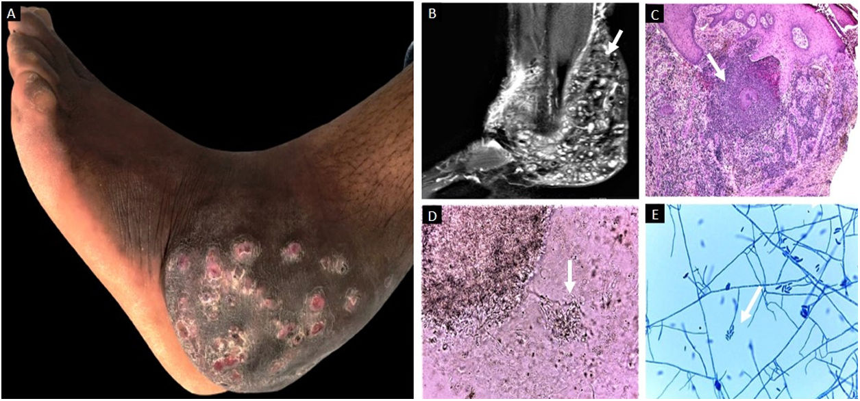
A 35-year-old man from São Tomé e Príncipe presented with a 12-year history of a slowly progressive left foot mass. Physical examination displayed an extensive unpainful and well-defined mass in his left ankle's posterolateral side. The lesion had multiple red, subcutaneous nodules with draining sinus tracts and grain discharge (Fig. 1A). Magnetic resonance imaging did not reveal osteomyelitis (Fig. 1B). A skin biopsy revealed granules in suppuration areas surrounded by eosinophilic material (Splendore-Hoeppli phenomenon) (Fig. 1C). Direct examination of the sinuses discharge showed multiple white grains with thin, septate hyphae (Fig. 1D). Microscopic examination of cultures revealed short conidiophores with macro-and microconidia (Fig. 1E). These findings support the diagnosis of white grain eumycetoma. The causative agent identified by polymerase chain reaction and DNA sequencing was Fusarium solani var keratoplasticum. The patient was treated with 400mg daily oral itraconazole for 24 months, with no response. Therapy was changed to 400mg daily oral voriconazole for 12 months, and he was referred to concomitant surgical excision. Eumycetoma, a neglected tropical disease, is a chronic and slowly progressive subcutaneous infection caused by several hyaline and dematiaceous fungi types. However, the genus Fusarium is a rare cause of mycetoma. Early recognition and treatment of this entity are essential.
Share