


A 32-year-old healthy Caucasian woman presented with a solitary, asymptomatic lesion on the right anterior forearm. She had first noticed this lesion 4 years earlier and hadn¿t related with nothing in particular; she also denied subsequent changes in size or appearance. She had no history of personal or family diseases.
Physical examinationPhysical examination revealed an oval, pink to brown, smooth yet finely wrinkled plaque, measuring 12x6mm (Fig. 1). Palpation was painless and the lesion didn¿t disappear with vitropression.
Histopathology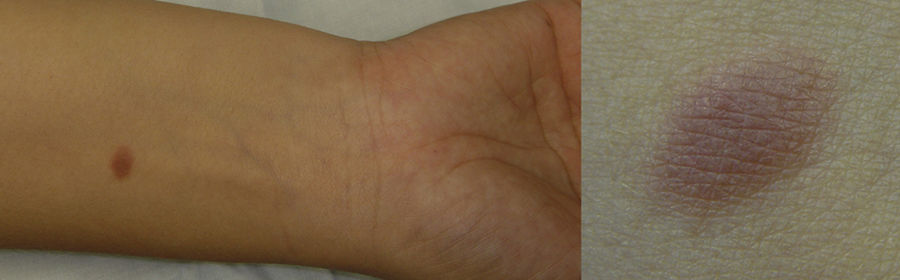
The histopathologic examination of an excisional biopsy showed a band like proliferation of fusiform cells, in a parallel distribution, seated on the papillary and upper reticular dermis, with no spreading to the subcutaneous tissue. Mitoses were rare and adnexal structures, nerves, and vessels were preserved (Fig. 2). Immunohistochemical staining demonstrated that these cells were CD34 positive, and factor XIIIa and S100 negative (Fig. 3).
Diagnosis

Taken together, the morphologic and immunophenotypic findings were felt to be most consistent with plaque-like CD34-positive dermal fibroma.
Course and treatmentExcisional biopsy was the offered treatment without any complication.
CommentMedallion-like dermal dendrocyte hamartoma (MDDH) also known as plaque-like CD34-positive dermal fibroma (PDF) was first described by Rodriguez-Jurado et al in 2004.1 The initial doubling of terminology has been elucidated as studies have suggested that the lesion neither is a hamartoma nor possess dermal dendrocytic differentiation. For these reasons, Kutzner and colleagues have suggested an adjustment in nomenclature to PDF, widening the spectrum of this tumor.2
PDF is a rare, benign, spindle cell neoplasm that classically presents as a congenital, asymptomatic, solitary, round- or triangular-shaped patch/plaque, on the neck or trunk, and most commonly in females.3,4 The overlying skin may be pink to brown, finely wrinkled, and/or atrophic.5 With the new nomenclature other cases were described, and this description has included congenital and acquired tumors, lesions located in different body sites rather than neck or trunk and also lacking the typical presentation. In fact, there are reports of lesions located on the limbs, hands and feet and also cases of indurated and angiomatous plaques, dermal nodules and pedunculated lesions.4,5 Our case represents an acquired form, however clinically very similar to the classical presentation.
Given its clinical heterogeneity, PDF may be clinically misdiagnosed as: congenital nevus; fibroblastic connective tissue nevus; neurofibroma; dermatofibroma; dermatomyofibroma; anetoderma; aplasia cutis; or, most importantly, as dermatofibrosarcoma protuberans (DFSP). It is critical to make the distinction between DFSP and PDF to prevent unnecessary wide excisions.4
Histology is characterized by a band-like dermal proliferation of spindle cells, which strongly express CD34. For this reason, the major differential diagnosis is with DFSP. Thus, it is important to fully characterize its histological and immunohistochemical findings and sometimes add cytogenetic and molecular studies when face with difficult or borderline cases.5 Immunohistochemically, the neoplastic cells are positive for CD34 and show variable expression of factor XIIIa and are negative for melanocytic markers (S100, MelanA, HMB45).3
We feel our case description is another example of a PDF. This case included, 21 occurrences of this rare neoplasm have been reported so far. Despite their histopathologic similarities, they have revealed an appreciable variability in clinical features. These differences have been related to the onset of the lesion (congenital or acquired), patient age at presentation, gender, location and specific clinical characteristics, and positivity to Factor XIIIa. Bearing this in mind, it is important to determine whether all apparent clinical variants are distinct subtypes of one same neoplasm or rather represent unique CD34-positive spindle cell proliferations.4
Although a spectrum of this entity is still being defined, PDF should always be considered within the differential diagnosis of CD34-positive dermal spindle cell neoplasms.3
Please cite this article as: Campos S, João A, Lencastre A. Placa de color rosa en superficie anterior de antebrazo derecho. Actas Dermosifiliogr. 2018;109:823–824.


