


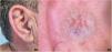

A 58-year-old man with no relevant past medical history presented with a lesion on the antihelix of the right ear (Fig. 1A). He had noted it for 6 years, during which it had grown slowly. He denied pain or any associated symptoms.
Physical examination
Examination revealed a well-circumscribed, firm, violaceous nodule measuring 1cm in diameter (Fig. 1A). Dermoscopy showed irregular, reticular-appearing linear vascular structures on an erythematous background (Fig. 1B). During surgery, the nodule was found to be solid and pseudoencapsulated and was submitted for histopathology.
HistopathologyHistology demonstrated a well-circumscribed tumor composed of smooth muscle fascicles encircling well-defined, thick-walled capillaries with semipermeable lumina (Fig. 2A–C). Immunohistochemistry tested positive for smooth muscle markers – actin (Fig. 2D), desmin, and calponin – and for the endothelial marker CD34 (Fig. 2E).

What is your diagnosis?
DiagnosisAngioleiomyoma.
Course and treatmentNo recurrence was observed 4 months after surgical excision with clear margins.
CommentAngioleiomyoma (ALM) is a usually benign cutaneous tumor derived from the smooth muscle layer in the tunica media of blood vessels. It accounts for 5% of all benign soft-tissue tumors and typically develops in the fifth to sixth decades of life. Although most commonly arising on the lower extremities, ALM can also occur on the head and neck. Pain is the most frequent presenting complaint; however, lesions on the head and neck are often asymptomatic.1–3
Clinically, ALM most often presents as a solitary, rounded, violaceous nodule with slow growth over years.4,5 Differential diagnosis includes purplish vascular lesions such as auricular hemangioma, glomus tumor, angiolipoma, angiolymphoid hyperplasia with eosinophilia, and the malignant variant of ALM, angioleiomyosarcoma. Cystic-appearing lesions such as epidermal cyst and auricular pseudocyst should also be considered.6,7 On dermoscopy, the violaceous hue reflects dilation of superficial vascular plexuses supplying the auricular cartilage.8,9 Definitive diagnosis is histologic, revealing interlacing smooth muscle fascicles enveloping blood vessels. Based on vessel composition and luminal caliber, ALM is categorized into capillary, venous, and cavernous subtypes.5,10,11 Immunohistochemistry is typically positive for vimentin, desmin, and actin, highlighting smooth muscle cell structures.12,13 ALM rarely recurs after excision; when recurrence occurs, the likelihood of malignancy is higher.14,15
Over the last 20 years, 15 case reports of auricular ALM have been published (Appendix A).16 There is a clear male predominance (14/15). Age ranged from 12 to 85 years; most patients (9/15) were in the 4th to 6th decades, 3 were younger than 30 years, and 3 were older than 60 years. In 11 cases, the lesion had been present for at least 1 year, and in 4 of those for more than 5 years. Only 2 of 15 cases reported tenderness or pain; the remainder were asymptomatic. Histologically, 13 of 15 were the venous subtype; the other 2 showed cavernous morphology. All cases were treated with surgical excision. Among those with clinical follow-up, no tumor recurrences were documented.
Conflicts of interestNone declared.
The following are the supplementary data to this article:


