array:24 [
  "pii" => "S0001731024004356"
  "issn" => "00017310"
  "doi" => "10.1016/j.ad.2024.05.014"
  "estado" => "S300"
  "fechaPublicacion" => "2024-07-01"
  "aid" => "3974"
  "copyright" => "AEDV"
  "copyrightAnyo" => "2024"
  "documento" => "simple-article"
  "crossmark" => 1
  "subdocumento" => "crp"
  "cita" => "Actas Dermosifiliogr. 2024;115:T732-T733"
  "abierto" => array:3 [
    "ES" => true
    "ES2" => true
    "LATM" => true
  ]
  "gratuito" => true
  "lecturas" => array:1 [
    "total" => 0
  ]
  "itemSiguiente" => array:18 [
    "pii" => "S0001731024002746"
    "issn" => "00017310"
    "doi" => "10.1016/j.ad.2023.06.025"
    "estado" => "S300"
    "fechaPublicacion" => "2024-07-01"
    "aid" => "3923"
    "copyright" => "AEDV"
    "documento" => "article"
    "crossmark" => 1
    "subdocumento" => "sco"
    "cita" => "Actas Dermosifiliogr. 2024;115:734-6"
    "abierto" => array:3 [
      "ES" => true
      "ES2" => true
      "LATM" => true
    ]
    "gratuito" => true
    "lecturas" => array:1 [
      "total" => 0
    ]
    "es" => array:10 [
      "idiomaDefecto" => true
      "cabecera" => "<span class="elsevierStyleTextfn">FORO DE RESIDENTES</span>"
      "titulo" => "FR-El papel de la dieta en el acn&#233;"
      "tienePdf" => "es"
      "tieneTextoCompleto" => "es"
      "paginas" => array:1 [
        0 => array:2 [
          "paginaInicial" => "734"
          "paginaFinal" => "736"
        ]
      ]
      "titulosAlternativos" => array:1 [
        "en" => array:1 [
          "titulo" => "RF-The role of diet in the management of acne"
        ]
      ]
      "contieneTextoCompleto" => array:1 [
        "es" => true
      ]
      "contienePdf" => array:1 [
        "es" => true
      ]
      "autores" => array:1 [
        0 => array:2 [
          "autoresLista" => "M&#46; Luque-Luna, M&#46; Sidro-Sarto"
          "autores" => array:2 [
            0 => array:2 [
              "nombre" => "M&#46;"
              "apellidos" => "Luque-Luna"
            ]
            1 => array:2 [
              "nombre" => "M&#46;"
              "apellidos" => "Sidro-Sarto"
            ]
          ]
        ]
      ]
    ]
    "idiomaDefecto" => "es"
    "EPUB" => "https://multimedia.elsevier.es/PublicationsMultimediaV1/item/epub/S0001731024002746?idApp=UINPBA000044"
    "url" => "/00017310/0000011500000007/v1_202407021756/S0001731024002746/v1_202407021756/es/main.assets"
  ]
  "itemAnterior" => array:18 [
    "pii" => "S0001731024002588"
    "issn" => "00017310"
    "doi" => "10.1016/j.ad.2023.04.047"
    "estado" => "S300"
    "fechaPublicacion" => "2024-07-01"
    "aid" => "3907"
    "copyright" => "AEDV"
    "documento" => "simple-article"
    "crossmark" => 1
    "subdocumento" => "crp"
    "cita" => "Actas Dermosifiliogr. 2024;115:732-3"
    "abierto" => array:3 [
      "ES" => true
      "ES2" => true
      "LATM" => true
    ]
    "gratuito" => true
    "lecturas" => array:1 [
      "total" => 0
    ]
    "es" => array:11 [
      "idiomaDefecto" => true
      "cabecera" => "<span class="elsevierStyleTextfn">DERMATOSCOPIA PR&#193;CTICA</span>"
      "titulo" => "Lo que la arruga esconde"
      "tienePdf" => "es"
      "tieneTextoCompleto" => "es"
      "paginas" => array:1 [
        0 => array:2 [
          "paginaInicial" => "732"
          "paginaFinal" => "733"
        ]
      ]
      "titulosAlternativos" => array:1 [
        "en" => array:1 [
          "titulo" => "What Wrinkles Actually Hide"
        ]
      ]
      "contieneTextoCompleto" => array:1 [
        "es" => true
      ]
      "contienePdf" => array:1 [
        "es" => true
      ]
      "resumenGrafico" => array:2 [
        "original" => 0
        "multimedia" => array:7 [
          "identificador" => "fig0005"
          "etiqueta" => "Figura 1"
          "tipo" => "MULTIMEDIAFIGURA"
          "mostrarFloat" => true
          "mostrarDisplay" => false
          "figura" => array:1 [
            0 => array:4 [
              "imagen" => "gr1.jpeg"
              "Alto" => 2258
              "Ancho" => 1508
              "Tamanyo" => 489255
            ]
          ]
          "descripcion" => array:1 [
            "es" => "<p id="spar0005" class="elsevierStyleSimplePara elsevierViewall">Presentaci&#243;n cl&#237;nica del caso&#58; zona cervical izquierda&#46;</p>"
          ]
        ]
      ]
      "autores" => array:1 [
        0 => array:2 [
          "autoresLista" => "G&#46; Baeza-Hern&#225;ndez, S&#46;P&#46; Herrero-Ruiz, A&#46;A&#46; Garrido-R&#237;os"
          "autores" => array:3 [
            0 => array:2 [
              "nombre" => "G&#46;"
              "apellidos" => "Baeza-Hern&#225;ndez"
            ]
            1 => array:2 [
              "nombre" => "S&#46;P&#46;"
              "apellidos" => "Herrero-Ruiz"
            ]
            2 => array:2 [
              "nombre" => "A&#46;A&#46;"
              "apellidos" => "Garrido-R&#237;os"
            ]
          ]
        ]
      ]
    ]
    "idiomaDefecto" => "es"
    "EPUB" => "https://multimedia.elsevier.es/PublicationsMultimediaV1/item/epub/S0001731024002588?idApp=UINPBA000044"
    "url" => "/00017310/0000011500000007/v1_202407021756/S0001731024002588/v1_202407021756/es/main.assets"
  ]
  "asociados" => array:1 [
    0 => array:18 [
      "pii" => "S0001731024002588"
      "issn" => "00017310"
      "doi" => "10.1016/j.ad.2023.04.047"
      "estado" => "S300"
      "fechaPublicacion" => "2024-07-01"
      "aid" => "3907"
      "copyright" => "AEDV"
      "documento" => "simple-article"
      "crossmark" => 1
      "subdocumento" => "crp"
      "cita" => "Actas Dermosifiliogr. 2024;115:732-3"
      "abierto" => array:3 [
        "ES" => true
        "ES2" => true
        "LATM" => true
      ]
      "gratuito" => true
      "lecturas" => array:1 [
        "total" => 0
      ]
      "es" => array:11 [
        "idiomaDefecto" => true
        "cabecera" => "<span class="elsevierStyleTextfn">DERMATOSCOPIA PR&#193;CTICA</span>"
        "titulo" => "Lo que la arruga esconde"
        "tienePdf" => "es"
        "tieneTextoCompleto" => "es"
        "paginas" => array:1 [
          0 => array:2 [
            "paginaInicial" => "732"
            "paginaFinal" => "733"
          ]
        ]
        "titulosAlternativos" => array:1 [
          "en" => array:1 [
            "titulo" => "What Wrinkles Actually Hide"
          ]
        ]
        "contieneTextoCompleto" => array:1 [
          "es" => true
        ]
        "contienePdf" => array:1 [
          "es" => true
        ]
        "resumenGrafico" => array:2 [
          "original" => 0
          "multimedia" => array:7 [
            "identificador" => "fig0005"
            "etiqueta" => "Figura 1"
            "tipo" => "MULTIMEDIAFIGURA"
            "mostrarFloat" => true
            "mostrarDisplay" => false
            "figura" => array:1 [
              0 => array:4 [
                "imagen" => "gr1.jpeg"
                "Alto" => 2258
                "Ancho" => 1508
                "Tamanyo" => 489255
              ]
            ]
            "descripcion" => array:1 [
              "es" => "<p id="spar0005" class="elsevierStyleSimplePara elsevierViewall">Presentaci&#243;n cl&#237;nica del caso&#58; zona cervical izquierda&#46;</p>"
            ]
          ]
        ]
        "autores" => array:1 [
          0 => array:2 [
            "autoresLista" => "G&#46; Baeza-Hern&#225;ndez, S&#46;P&#46; Herrero-Ruiz, A&#46;A&#46; Garrido-R&#237;os"
            "autores" => array:3 [
              0 => array:2 [
                "nombre" => "G&#46;"
                "apellidos" => "Baeza-Hern&#225;ndez"
              ]
              1 => array:2 [
                "nombre" => "S&#46;P&#46;"
                "apellidos" => "Herrero-Ruiz"
              ]
              2 => array:2 [
                "nombre" => "A&#46;A&#46;"
                "apellidos" => "Garrido-R&#237;os"
              ]
            ]
          ]
        ]
      ]
      "idiomaDefecto" => "es"
      "EPUB" => "https://multimedia.elsevier.es/PublicationsMultimediaV1/item/epub/S0001731024002588?idApp=UINPBA000044"
      "url" => "/00017310/0000011500000007/v1_202407021756/S0001731024002588/v1_202407021756/es/main.assets"
    ]
  ]
  "en" => array:14 [
    "idiomaDefecto" => true
    "cabecera" => "<span class="elsevierStyleTextfn">Practical Dermoscopy</span>"
    "titulo" => " What Wrinkles Actually Hide"
    "tieneTextoCompleto" => true
    "paginas" => array:1 [
      0 => array:2 [
        "paginaInicial" => "T732"
        "paginaFinal" => "T733"
      ]
    ]
    "autores" => array:1 [
      0 => array:4 [
        "autoresLista" => "G&#46; Baeza-Hern&#225;ndez, S&#46;P&#46; Herrero-Ruiz, A&#46;A&#46; Garrido-R&#237;os"
        "autores" => array:3 [
          0 => array:4 [
            "nombre" => "G&#46;"
            "apellidos" => "Baeza-Hern&#225;ndez"
            "email" => array:1 [
              0 => "gloria.baezahdez@outlook.com"
            ]
            "referencia" => array:1 [
              0 => array:2 [
                "etiqueta" => "<span class="elsevierStyleSup">&#42;</span>"
                "identificador" => "cor0005"
              ]
            ]
          ]
          1 => array:2 [
            "nombre" => "S&#46;P&#46;"
            "apellidos" => "Herrero-Ruiz"
          ]
          2 => array:2 [
            "nombre" => "A&#46;A&#46;"
            "apellidos" => "Garrido-R&#237;os"
          ]
        ]
        "afiliaciones" => array:1 [
          0 => array:2 [
            "entidad" => "Servicio de Dermatolog&#237;a&#44; Hospital Universitario de Fuenlabrada&#44; Madrid&#44; Spain"
            "identificador" => "aff0005"
          ]
        ]
        "correspondencia" => array:1 [
          0 => array:3 [
            "identificador" => "cor0005"
            "etiqueta" => "&#8270;"
            "correspondencia" => "Corresponding author&#46;"
          ]
        ]
      ]
    ]
    "titulosAlternativos" => array:1 [
      "es" => array:1 [
        "titulo" => "Lo que la arruga esconde"
      ]
    ]
    "resumenGrafico" => array:2 [
      "original" => 0
      "multimedia" => array:7 [
        "identificador" => "fig0010"
        "etiqueta" => "Figure 2"
        "tipo" => "MULTIMEDIAFIGURA"
        "mostrarFloat" => true
        "mostrarDisplay" => false
        "figura" => array:1 [
          0 => array:4 [
            "imagen" => "gr2.jpeg"
            "Alto" => 1188
            "Ancho" => 900
            "Tamanyo" => 113057
          ]
        ]
        "descripcion" => array:1 [
          "en" => "<p id="spar0010" class="elsevierStyleSimplePara elsevierViewall">Dermoscopic images with polarized light &#40;a and b&#41;&#46; Absence of pigmented network&#46; Presence of maple leaf-like structures&#44; dots&#44; blue-gray globules&#44; ulceration&#44; and several erosions&#46;</p>"
        ]
      ]
    ]
    "textoCompleto" => "<span class="elsevierStyleSections"><span id="sec0005" class="elsevierStyleSection elsevierViewall"><span class="elsevierStyleSectionTitle" id="sect0005">Case presentation</span><p id="par0005" class="elsevierStylePara elsevierViewall">A 70-year-old woman&#44; with no relevant past medical history&#44; presented with a 1-year history asymptomatic lesion on her neck&#46; Upon examination&#44; she showed a 30<span class="elsevierStyleHsp" style=""></span>mm x 2<span class="elsevierStyleHsp" style=""></span>mm linear erythematous-brownish plaque&#44; somewhat crusted&#44; located in the depth of a fold on the left side of her neck &#40;<a class="elsevierStyleCrossRef" href="#fig0005">Fig&#46; 1</a>&#41;&#46;</p><elsevierMultimedia ident="fig0005"></elsevierMultimedia><p id="par0010" class="elsevierStylePara elsevierViewall">What is your diagnosis&#63;</p></span><span id="sec0010" class="elsevierStyleSection elsevierViewall"><span class="elsevierStyleSectionTitle" id="sect0010">Comment</span><p id="par0015" class="elsevierStylePara elsevierViewall">Dermoscopic findings shown in <a class="elsevierStyleCrossRef" href="#fig0010">Fig&#46; 2</a> are consistent with a non-superficial basal cell carcinoma&#46; Complete excision of the lesion was performed&#44; which confirmed the diagnosis of nodular and micronodular basal cell carcinoma with clear margins&#46;</p><elsevierMultimedia ident="fig0010"></elsevierMultimedia><p id="par0020" class="elsevierStylePara elsevierViewall">In our case&#44; clinical differential diagnosis also included the following entities&#58; scar&#44; exogenous pigmentation&#44; inverse pigmented lichen planus&#44; and terra firma-forme dermatosis&#46; Dermoscopy allowed us to establish an early diagnosis of basal cell carcinoma&#46;</p><p id="par0025" class="elsevierStylePara elsevierViewall">Linear basal cell carcinoma was first described in 1985<a class="elsevierStyleCrossRef" href="#bib0035"><span class="elsevierStyleSup">1</span></a> and is most commonly located in the periocular area and the neck<a class="elsevierStyleCrossRefs" href="#bib0040"><span class="elsevierStyleSup">2&#8211;6</span></a>&#59; the pathogenic mechanism by which the tumor tends to grow along skin tension lines is unknown to this date&#46;<a class="elsevierStyleCrossRefs" href="#bib0040"><span class="elsevierStyleSup">2&#44;3</span></a> No specific dermoscopic findings have ever been described for the linear variant of basal cell carcinoma&#46;<a class="elsevierStyleCrossRefs" href="#bib0040"><span class="elsevierStyleSup">2&#44;4&#44;5</span></a> A recently published retrospective&#44; descriptive study with 18 tumors of this type has analyzed their dermoscopic features<a class="elsevierStyleCrossRef" href="#bib0050"><span class="elsevierStyleSup">4</span></a>&#58; more than 80&#37; were pigmented&#44; as in our case&#44; a higher proportion than expected perhaps due to the ethnic characteristics of the study patients &#40;mostly Hispanic&#47;Latino&#41; and a difficulty in diagnosing non-pigmented linear basal cell carcinoma that may lead to underdiagnosis&#46;<a class="elsevierStyleCrossRefs" href="#bib0050"><span class="elsevierStyleSup">4&#44;5</span></a> Dermoscopic structures specific to basal cell carcinoma most frequently observed were blue-gray globules&#44; well-focused dots&#44; and maple leaf-like structures<a class="elsevierStyleCrossRef" href="#bib0050"><span class="elsevierStyleSup">4</span></a> &#40;all present in our case&#41;&#46;</p><p id="par0030" class="elsevierStylePara elsevierViewall">We wish to mention the utility of dermoscopy to diagnose linear basal cell carcinoma&#44; exclude other diseases that may also present a linear morphology&#44;<a class="elsevierStyleCrossRef" href="#bib0050"><span class="elsevierStyleSup">4</span></a> and more accurately delineate the tumor&#39;s extension&#44; as reportedly&#44; tumors have a greater-than-expected subclinical extension&#46;<a class="elsevierStyleCrossRefs" href="#bib0045"><span class="elsevierStyleSup">3&#44;6</span></a> Furthermore&#44; dermoscopy can help guide the histological subtype before anatomopathological results become available and classify it as low or high risk&#44; thus allowing for the selection of the most appropriate surgical technique and margins&#46;<a class="elsevierStyleCrossRefs" href="#bib0040"><span class="elsevierStyleSup">2&#44;6</span></a> The most common histological subtype of linear basal cell carcinoma is nodular&#44;<a class="elsevierStyleCrossRefs" href="#bib0045"><span class="elsevierStyleSup">3&#8211;6</span></a> although it can sometimes be associated with more aggressive subtypes&#44; such as the infiltrative or morpheaform subtypes&#46;<a class="elsevierStyleCrossRefs" href="#bib0045"><span class="elsevierStyleSup">3&#44;4</span></a> In retrospective studies and case series&#44; Mohs surgery is the most widely used treatment for linear basal cell carcinoma&#44;<a class="elsevierStyleCrossRefs" href="#bib0045"><span class="elsevierStyleSup">3&#44;4&#44;6</span></a> although other authors have also achieved good results with conventional surgery&#44; as in our case&#44; and recommend reserving Mohs surgery for aggressive histological subtypes&#44; or those located in high or medium risk areas &#40;H or M areas&#41;&#46;<a class="elsevierStyleCrossRef" href="#bib0060"><span class="elsevierStyleSup">6</span></a></p></span><span id="sec0015" class="elsevierStyleSection elsevierViewall"><span class="elsevierStyleSectionTitle" id="sect0015">Conflict of interests</span><p id="par0035" class="elsevierStylePara elsevierViewall">The authors state that they have no conflict of interests&#46;</p></span></span>"
    "textoCompletoSecciones" => array:1 [
      "secciones" => array:4 [
        0 => array:2 [
          "identificador" => "sec0005"
          "titulo" => "Case presentation"
        ]
        1 => array:2 [
          "identificador" => "sec0010"
          "titulo" => "Comment"
        ]
        2 => array:2 [
          "identificador" => "sec0015"
          "titulo" => "Conflict of interests"
        ]
        3 => array:1 [
          "titulo" => "References"
        ]
      ]
    ]
    "pdfFichero" => "main.pdf"
    "tienePdf" => true
    "multimedia" => array:2 [
      0 => array:7 [
        "identificador" => "fig0005"
        "etiqueta" => "Figure 1"
        "tipo" => "MULTIMEDIAFIGURA"
        "mostrarFloat" => true
        "mostrarDisplay" => false
        "figura" => array:1 [
          0 => array:4 [
            "imagen" => "gr1.jpeg"
            "Alto" => 897
            "Ancho" => 600
            "Tamanyo" => 87964
          ]
        ]
        "descripcion" => array:1 [
          "en" => "<p id="spar0005" class="elsevierStyleSimplePara elsevierViewall">Clinical case presentation&#58; left cervical area&#46;</p>"
        ]
      ]
      1 => array:7 [
        "identificador" => "fig0010"
        "etiqueta" => "Figure 2"
        "tipo" => "MULTIMEDIAFIGURA"
        "mostrarFloat" => true
        "mostrarDisplay" => false
        "figura" => array:1 [
          0 => array:4 [
            "imagen" => "gr2.jpeg"
            "Alto" => 1188
            "Ancho" => 900
            "Tamanyo" => 113057
          ]
        ]
        "descripcion" => array:1 [
          "en" => "<p id="spar0010" class="elsevierStyleSimplePara elsevierViewall">Dermoscopic images with polarized light &#40;a and b&#41;&#46; Absence of pigmented network&#46; Presence of maple leaf-like structures&#44; dots&#44; blue-gray globules&#44; ulceration&#44; and several erosions&#46;</p>"
        ]
      ]
    ]
    "bibliografia" => array:2 [
      "titulo" => "References"
      "seccion" => array:1 [
        0 => array:2 [
          "identificador" => "bibs0015"
          "bibliografiaReferencia" => array:6 [
            0 => array:3 [
              "identificador" => "bib0035"
              "etiqueta" => "1"
              "referencia" => array:1 [
                0 => array:2 [
                  "contribucion" => array:1 [
                    0 => array:2 [
                      "titulo" => "Linear basal cell epithelioma"
                      "autores" => array:1 [
                        0 => array:2 [
                          "etal" => false
                          "autores" => array:1 [
                            0 => "J&#46;E&#46; Lewis"
                          ]
                        ]
                      ]
                    ]
                  ]
                  "host" => array:1 [
                    0 => array:2 [
                      "doi" => "10.1111/j.1365-4362.1985.tb05396.x"
                      "Revista" => array:6 [
                        "tituloSerie" => "Int J Dermatol&#46;"
                        "fecha" => "1985"
                        "volumen" => "24"
                        "paginaInicial" => "124"
                        "paginaFinal" => "125"
                        "link" => array:1 [
                          0 => array:2 [
                            "url" => "https://www.ncbi.nlm.nih.gov/pubmed/3988420"
                            "web" => "Medline"
                          ]
                        ]
                      ]
                    ]
                  ]
                ]
              ]
            ]
            1 => array:3 [
              "identificador" => "bib0040"
              "etiqueta" => "2"
              "referencia" => array:1 [
                0 => array:2 [
                  "contribucion" => array:1 [
                    0 => array:2 [
                      "titulo" => "Dermatoscopia del carcinoma basocelular&#58; revisi&#243;n actualizada"
                      "autores" => array:1 [
                        0 => array:2 [
                          "etal" => false
                          "autores" => array:3 [
                            0 => "M&#46; &#193;lvarez-Salafranca"
                            1 => "M&#46; Ara"
                            2 => "P&#46; Zaballos"
                          ]
                        ]
                      ]
                    ]
                  ]
                  "host" => array:1 [
                    0 => array:2 [
                      "doi" => "10.1016/j.ad.2020.11.011"
                      "Revista" => array:5 [
                        "tituloSerie" => "Actas Dermosifiliogr&#46;"
                        "fecha" => "2021"
                        "volumen" => "112"
                        "paginaInicial" => "330"
                        "paginaFinal" => "338"
                      ]
                    ]
                  ]
                ]
              ]
            ]
            2 => array:3 [
              "identificador" => "bib0045"
              "etiqueta" => "3"
              "referencia" => array:1 [
                0 => array:2 [
                  "contribucion" => array:1 [
                    0 => array:2 [
                      "titulo" => "Linear basal cell carcinoma&#58; A distinct condition&#63;"
                      "autores" => array:1 [
                        0 => array:2 [
                          "etal" => false
                          "autores" => array:2 [
                            0 => "F&#46; Al-Niaimi"
                            1 => "C&#46;C&#46; Lyon"
                          ]
                        ]
                      ]
                    ]
                  ]
                  "host" => array:1 [
                    0 => array:2 [
                      "doi" => "10.1111/j.1365-2230.2010.03908.x"
                      "Revista" => array:6 [
                        "tituloSerie" => "Clin Exp Dermatol&#46;"
                        "fecha" => "2011"
                        "volumen" => "36"
                        "paginaInicial" => "231"
                        "paginaFinal" => "234"
                        "link" => array:1 [
                          0 => array:2 [
                            "url" => "https://www.ncbi.nlm.nih.gov/pubmed/20659116"
                            "web" => "Medline"
                          ]
                        ]
                      ]
                    ]
                  ]
                ]
              ]
            ]
            3 => array:3 [
              "identificador" => "bib0050"
              "etiqueta" => "4"
              "referencia" => array:1 [
                0 => array:2 [
                  "contribucion" => array:1 [
                    0 => array:2 [
                      "titulo" => "Dermoscopy of linear basal cell carcinomas&#44; a potential mimicker of linear lesions&#58; a descriptive case series"
                      "autores" => array:1 [
                        0 => array:2 [
                          "etal" => true
                          "autores" => array:6 [
                            0 => "C&#46; Navarrete-Dechent"
                            1 => "M&#46;A&#46; Marchetti"
                            2 => "P&#46; Uribe"
                            3 => "R&#46;J&#46; Schwartz"
                            4 => "K&#46; Liopyris"
                            5 => "N&#46;G&#46; Marghoob"
                          ]
                        ]
                      ]
                    ]
                  ]
                  "host" => array:1 [
                    0 => array:2 [
                      "doi" => "10.5826/dpc.1204a19"
                      "Revista" => array:5 [
                        "tituloSerie" => "Dermatol Pract Concept&#46;"
                        "fecha" => "2022"
                        "volumen" => "12"
                        "paginaInicial" => "e2022195"
                        "link" => array:1 [
                          0 => array:2 [
                            "url" => "https://www.ncbi.nlm.nih.gov/pubmed/36534556"
                            "web" => "Medline"
                          ]
                        ]
                      ]
                    ]
                  ]
                ]
              ]
            ]
            4 => array:3 [
              "identificador" => "bib0055"
              "etiqueta" => "5"
              "referencia" => array:1 [
                0 => array:2 [
                  "contribucion" => array:1 [
                    0 => array:2 [
                      "titulo" => "Linear basal cell carcinoma&#58; Report of three cases with dermoscopic findings"
                      "autores" => array:1 [
                        0 => array:2 [
                          "etal" => false
                          "autores" => array:4 [
                            0 => "C&#46;M&#46; Alc&#225;ntara-Reifs"
                            1 => "R&#46; Salido-Vallejo"
                            2 => "A&#46; Gonz&#225;lez-Menchen"
                            3 => "A&#46; V&#233;lez Garc&#237;a-Nieto"
                          ]
                        ]
                      ]
                    ]
                  ]
                  "host" => array:1 [
                    0 => array:2 [
                      "doi" => "10.4103/0378-6323.190850"
                      "Revista" => array:5 [
                        "tituloSerie" => "Indian J Dermatol Venereol Leprol&#46;"
                        "fecha" => "2016"
                        "volumen" => "82"
                        "paginaInicial" => "708"
                        "paginaFinal" => "711"
                      ]
                    ]
                  ]
                ]
              ]
            ]
            5 => array:3 [
              "identificador" => "bib0060"
              "etiqueta" => "6"
              "referencia" => array:1 [
                0 => array:2 [
                  "contribucion" => array:1 [
                    0 => array:2 [
                      "titulo" => "Linear basal cell carcinoma occurs most commonly on the lower eyelid"
                      "autores" => array:1 [
                        0 => array:2 [
                          "etal" => false
                          "autores" => array:4 [
                            0 => "G&#46;L&#46; Becher"
                            1 => "A&#46; Affleck"
                            2 => "C&#46; Fleming"
                            3 => "A&#46; Evans"
                          ]
                        ]
                      ]
                    ]
                  ]
                  "host" => array:1 [
                    0 => array:2 [
                      "doi" => "10.1111/j.1365-2230.2010.03950.x"
                      "Revista" => array:5 [
                        "tituloSerie" => "Clin Exp Dermatol&#46;"
                        "fecha" => "2011"
                        "volumen" => "36"
                        "paginaInicial" => "311"
                        "paginaFinal" => "312"
                      ]
                    ]
                  ]
                ]
              ]
            ]
          ]
        ]
      ]
    ]
  ]
  "idiomaDefecto" => "en"
  "url" => "/00017310/0000011500000007/v1_202407021756/S0001731024004356/v1_202407021756/en/main.assets"
  "Apartado" => array:4 [
    "identificador" => "19290"
    "tipo" => "SECCION"
    "es" => array:2 [
      "titulo" => "Dermatoscopia pr&#225;ctica"
      "idiomaDefecto" => true
    ]
    "idiomaDefecto" => "es"
  ]
  "PDF" => "https://static.elsevier.es/multimedia/00017310/0000011500000007/v1_202407021756/S0001731024004356/v1_202407021756/en/main.pdf?idApp=UINPBA000044&text.app=https://actasdermo.org/"
  "EPUB" => "https://multimedia.elsevier.es/PublicationsMultimediaV1/item/epub/S0001731024004356?idApp=UINPBA000044"
]
Compartir
Información de la revista
Vol. 115. Núm. 7.
Páginas T732-T733 (julio - agosto 2024)
Compartir
Compartir
Descargar PDF
Más opciones de artículo
Visitas
1158
Vol. 115. Núm. 7.
Páginas T732-T733 (julio - agosto 2024)
Practical Dermoscopy
Acceso a texto completo
What Wrinkles Actually Hide
Lo que la arruga esconde
Visitas
1158
G. Baeza-Hernández
Autor para correspondencia
gloria.baezahdez@outlook.com

Corresponding author.
, S.P. Herrero-Ruiz, A.A. Garrido-Ríos
Servicio de Dermatología, Hospital Universitario de Fuenlabrada, Madrid, Spain
Contenido relacionado
Actas Dermosifiliogr. 2024;115:732-310.1016/j.ad.2023.04.047
G. Baeza-Hernández, S.P. Herrero-Ruiz, A.A. Garrido-Ríos
Este artículo ha recibido
Información del artículo
Texto completo
Bibliografía
Descargar PDF
Estadísticas
Figuras (2)
Texto completo
Case presentation

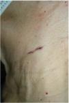
A 70-year-old woman, with no relevant past medical history, presented with a 1-year history asymptomatic lesion on her neck. Upon examination, she showed a 30mm x 2mm linear erythematous-brownish plaque, somewhat crusted, located in the depth of a fold on the left side of her neck (Fig. 1).

Figure 1.

Clinical case presentation: left cervical area.

(0.08MB).

What is your diagnosis?

Comment

Dermoscopic findings shown in Fig. 2 are consistent with a non-superficial basal cell carcinoma. Complete excision of the lesion was performed, which confirmed the diagnosis of nodular and micronodular basal cell carcinoma with clear margins.

Figure 2.

Dermoscopic images with polarized light (a and b). Absence of pigmented network. Presence of maple leaf-like structures, dots, blue-gray globules, ulceration, and several erosions.

(0.11MB).

In our case, clinical differential diagnosis also included the following entities: scar, exogenous pigmentation, inverse pigmented lichen planus, and terra firma-forme dermatosis. Dermoscopy allowed us to establish an early diagnosis of basal cell carcinoma.

Linear basal cell carcinoma was first described in 19851 and is most commonly located in the periocular area and the neck2–6; the pathogenic mechanism by which the tumor tends to grow along skin tension lines is unknown to this date.2,3 No specific dermoscopic findings have ever been described for the linear variant of basal cell carcinoma.2,4,5 A recently published retrospective, descriptive study with 18 tumors of this type has analyzed their dermoscopic features4: more than 80% were pigmented, as in our case, a higher proportion than expected perhaps due to the ethnic characteristics of the study patients (mostly Hispanic/Latino) and a difficulty in diagnosing non-pigmented linear basal cell carcinoma that may lead to underdiagnosis.4,5 Dermoscopic structures specific to basal cell carcinoma most frequently observed were blue-gray globules, well-focused dots, and maple leaf-like structures4 (all present in our case).

We wish to mention the utility of dermoscopy to diagnose linear basal cell carcinoma, exclude other diseases that may also present a linear morphology,4 and more accurately delineate the tumor's extension, as reportedly, tumors have a greater-than-expected subclinical extension.3,6 Furthermore, dermoscopy can help guide the histological subtype before anatomopathological results become available and classify it as low or high risk, thus allowing for the selection of the most appropriate surgical technique and margins.2,6 The most common histological subtype of linear basal cell carcinoma is nodular,3–6 although it can sometimes be associated with more aggressive subtypes, such as the infiltrative or morpheaform subtypes.3,4 In retrospective studies and case series, Mohs surgery is the most widely used treatment for linear basal cell carcinoma,3,4,6 although other authors have also achieved good results with conventional surgery, as in our case, and recommend reserving Mohs surgery for aggressive histological subtypes, or those located in high or medium risk areas (H or M areas).6

Conflict of interests

The authors state that they have no conflict of interests.

References
[1]
J.E. Lewis.
Linear basal cell epithelioma.
Int J Dermatol., 24 (1985), pp. 124-125
[2]
M. Álvarez-Salafranca, M. Ara, P. Zaballos.
Dermatoscopia del carcinoma basocelular: revisión actualizada.
Actas Dermosifiliogr., 112 (2021), pp. 330-338
[3]
F. Al-Niaimi, C.C. Lyon.
Linear basal cell carcinoma: A distinct condition?.
Clin Exp Dermatol., 36 (2011), pp. 231-234
[4]
C. Navarrete-Dechent, M.A. Marchetti, P. Uribe, R.J. Schwartz, K. Liopyris, N.G. Marghoob, et al.
Dermoscopy of linear basal cell carcinomas, a potential mimicker of linear lesions: a descriptive case series.
Dermatol Pract Concept., 12 (2022), pp. e2022195
[5]
C.M. Alcántara-Reifs, R. Salido-Vallejo, A. González-Menchen, A. Vélez García-Nieto.
Linear basal cell carcinoma: Report of three cases with dermoscopic findings.
Indian J Dermatol Venereol Leprol., 82 (2016), pp. 708-711
[6]
G.L. Becher, A. Affleck, C. Fleming, A. Evans.
Linear basal cell carcinoma occurs most commonly on the lower eyelid.
Clin Exp Dermatol., 36 (2011), pp. 311-312
Copyright © 2024. AEDV
Descargar PDF
Idiomas
Actas Dermo-Sifiliográficas
Opciones de artículo
Herramientas
es en

¿Es usted profesional sanitario apto para prescribir o dispensar medicamentos?

Are you a health professional able to prescribe or dispense drugs?