


A 21-year-old white man with no personal history of interest was referred from primary care for an asymptomatic lesion on the right arm that had appeared 10 months earlier.
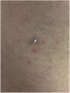
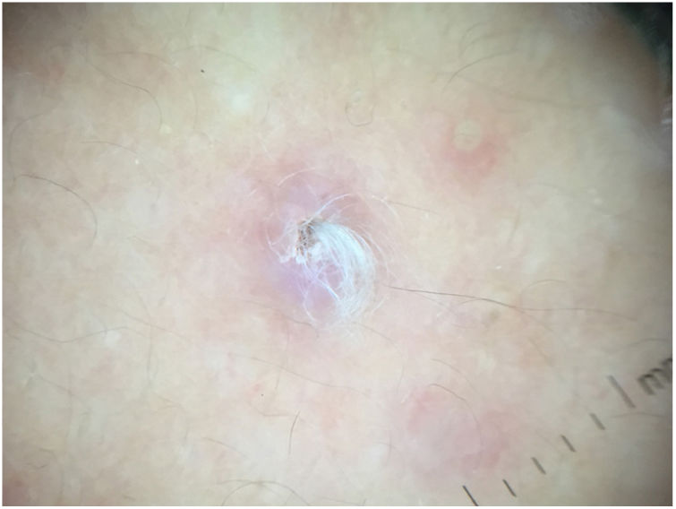
Physical ExaminationPhysical examination revealed a lesion of approximately 4mm in diameter, with a nodulocystic appearance and a central tuft of white hairs (Fig. 1). Dermoscopy showed a white–pink area with a slightly depressed and scaly center from which a tuft of fine whitish hair emerged. No indications of a melanocytic lesion nor any specific vascular pattern were observed (Fig. 2).
Histopathology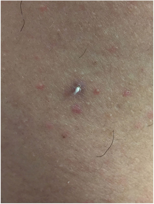
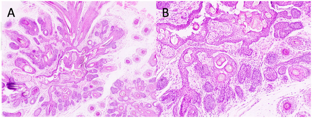
Biopsy after excision showed a dilated central follicle from which numerous small secondary and tertiary follicles arose, with panfollicular differentiation. The central follicle contained keratin and hair shafts. The small follicles were largely mature, and abortive follicles radiated into the surrounding dermis. Sebaceous glands were detected (Fig. 3).
What is your diagnosis?
DiagnosisTrichofolliculoma.
CommentTrichofolliculoma (TF) is a benign, infrequent hamartoma with follicular differentiation, and is more commonly found in adults. No sexual predilection has been described. It occurs spontaneously, although appearance after trauma has also been reported. It usually presents as a solitary, normal skin-colored papule of variable size, with a central depression from which a tuft of fine white hair emerges. The most common location is the face, followed by the neck and scalp.1,2 However, the literature includes cases of congenital TF, multiple TF, and extrafacial TF. The clinical differential diagnosis includes entities such as fibrous papule of the nose, fistula of the dorsum of the nose, and trichostasis spinulosa.
Histology is diagnostic and characteristic, revealing cyst-like dilation of the infundibulum, infundibular cornification, and central orthokeratin. Secondary and tertiary hair follicles radiate radially from this central structure. The perilesional stroma is fibrous and lamellar and is characteristically separated by clefts from the stroma of the adjacent healthy dermis. Piloerector muscles are usually absent and sebaceous differentiation is minimal in typical cases.3 Secondary follicles containing abundant sebaceous acini are a feature of a variant known as sebaceous TF. Several authors distinguish 3 stages of TF (early, complete or developed, and late) that correspond to the hair follicle cycle. The most striking sebaceous differentiation is observed in late TF. Some authors even argue that so-called folliculosebaceous cystic hamartoma may correspond to longstanding TF. However, according to other authors these are distinct entities.4 No cases of malignant transformation have been described to date, and published cases of TF with perineural infiltration appear to correspond more to microcystic adnexal carcinomas. The main histological differential diagnoses are dilated pore of Winer, fibrous papule of the nose, fibrofolliculoma, and hair follicle nevus.3
Descriptions of dermoscopic findings are scarce, and vary depending on the stage of evolution of the lesion. Panasti et al. described a “fireworks” pattern in a TF lesion of 4 months duration. This pattern consists of a central brown area with dark brown radial projections and the absence of a pigmented reticulum. García-García et al. recently described the following dermoscopic characteristics of a TF lesion that had appeared 1 year earlier: a bluish nodule with a central whitish–pink area, bright white structures, punctate vessels, and central scaling.5,6 This pattern more closely resembles that observed in our patient, whose lesion also featured a characteristic tuft of fine white hair. In summary, the role of dermoscopy in the diagnosis of adnexal tumors remains to be defined.
Because TF is a benign entity, treatment is not required, except for diagnostic or cosmetic reasons. In contrast to other pilar tumors, no associations with any major disease are described in the literature.
Conflicts of InterestThe authors declare that they have no conflicts of interest.


