Tissue expanders are temporary prostheses that progressively expand over time gradually increasing the surface area of tissue, making them useful in reconstructive surgery of soft parts.1
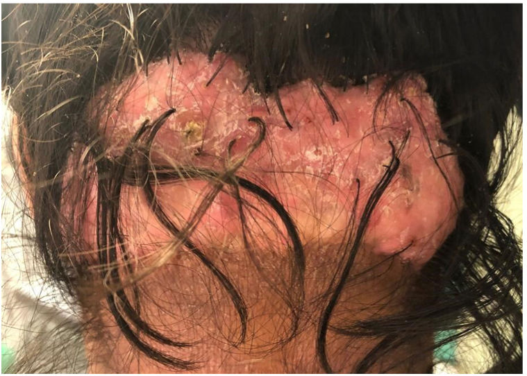
A 36-year-old woman, with a previous clinical diagnosis of acne keloidalis nuchae, presented with a 15-year history of skin lesion in the occipital region. After having followed several lines of medical therapy, such as antibiotics, topical, intralesional and oral corticosteroids, and oral retinoids she persistently exhibited a large 15cm x 5cm scarred, erythematous, tuberous lesion in the occipitonuchal region, with a scarred appearance and polytrichia (hair follicles in tuft) (Fig. 1).

Given the impossibility of a simple surgical treatment, several options were considered, such as a local flap, a microvascularized free flap, or tissue expansion. After discussion with the patient, we decided to use a tissue expander.
Description of the techniqueThe technique involves the gradual expansion of a silicone prosthesis implanted underneath the skin adjacent to the defect, leaving a 5cm margin of healthy skin to avoid expanding the affected skin. The expander is progressively filled with distilled water (this is an important step, because the use of a saline solution can cause precipitates that can later complicate emptying), through a self-sealing filling port. As the balloon increases in size, the tissue adapts thanks to its elastic properties, increasing its length and mass through mechanisms known as mechanical creep (stretching of collagen fibers) and biological creep (which stimulates the growth of new tissue).2
The procedure to remove the lesion with the expander requires 2 surgical interventions: the first one for placement and the second for removing the scar tissue and covering the defect with the expanded tissue.
In our case, during the first surgical intervention, the expander was placed between the subcutaneous cellular tissue and the galea, as the video shows. The tube that connected to the filling valve was, then, brought outwards through a small incision through which the expander was gradually filled.
After successive outpatient visits and gradual filling with a final volume of about 200mL of SSF throughout 6 months, the second surgical intervention was performed. The occipital lesion was excised through a simple excision and the defect was covered with an advancement flap from the region of the expander.
The aesthetic result was optimal after the surgical intervention, as the barely visible scar matched the posterior hairline, and there was no evidence of disease recurrence at the 1-year follow-up.
Indications- •
Removal of scars: mainly post-burn.
- •
Coverage of defects after removal of benign lesions: giant melanocytic nevus, sebaceous nevus, leishmaniasis...
- •
Reconstruction of defects in highly sensitive areas: face, scalp, auricle.
- •
Breast reconstruction.
- •
Coverage of pressure ulcers.
Although there are no absolute contraindications for the placement of a tissue expander, the degree of aesthetic deformity during the expansion process and risk factors for complications should be taken into consideration.
Risk factors associated with a higher rate of complications with the use of tissue expanders in reconstructive surgery are BMI > 30, history of radiotherapy, age older than 50 years, smoking, number of expanders placed, location in lower limbs, and size of the defect.
ComplicationsThe rate of complications ranges from 10% up to 20% depending on the series.3 The most common complications based on the stage of the procedure are:
- -
During the initial expander placement surgery: perforation of the skin overlying the pocket and trauma to important anatomical tissues (such as nerves, veins, arteries, fascia, or muscle).
- -
During the expansion: pocket perforation, prosthesis migration, infection, and necrosis of a portion of the expanded tissue due to excessive tension from fluid overinjection, or infection.
- -
During the second defect coverage surgery and thereafter: seroma of the expander capsule, hematoma, and necrosis of the distal portion of the flap.
Tissue expanders are a valuable option for reconstructing large surgical defects in aesthetically sensitive regions (such as the face or scalp) where primary closure via local skin flap is not possible. Moreover, the technique provides good aesthetic results since it preserves the same texture, color, appendages, and sensitivity characteristics as the excised tissue.
As disadvantages, we find the aesthetic deformity caused during the expansion of the prosthesis (whose filling time will depend on the size of the defect) and the need for 2 surgical interventions.
Tissue expansion procedure is a valuable adjuvant resource to treat many challenging cases that are not amenable to correction with traditional surgical techniques.
Conflicts of interestNone declared.


