


The Canary Islands (Spain) serve as the entry point to the European Union for thousands of people each year, many of whom arrive by sea in small boats known as “pateras”. The extreme conditions of the journey often result in complex clinical presentations upon arrival.
ObjectiveTo evaluate the characteristics of patients admitted to a tertiary referral center after a journey in a patera boat.
Materials and methodsWe conducted a retrospective cross-sectional observational study including migrant patients after a journey in a patera boat who required admission to CHUNSC during 2023. The study evaluates the care pathway and the patients’ epidemiological, clinical, and evolutionary characteristics.
ResultsA total of 484 patients were treated in the emergency department. A total of 93 (19.2%) required admission, the mean age was 23 years old, and 85 (91.4%) were men. The mean length of stay was 29 days. A total of 22 patients (23.6%) were admitted to the ICU for a mean 7.6 days. The most common clinical conditions upon admission were anemia (73.1%) and rhabdomyolysis (62.5%). A total of 76 patients exhibited skin lesions (81.7%), predominantly on the legs (68.4%), feet (67.1%), and buttocks and sacrum (56.6%). A total of 36 (38.7%) underwent surgery; debridement of skin necrosis was performed in 22, skin grafts in 21, and amputations in 12. A total of 35 positive cultures were obtained, with Staphylococcus aureus, Pseudomonas aeruginosa and Shewanella algae being the most frequently isolated pathogens.
ConclusionsThe extreme conditions of the journey in a patera lead to complex and severe clinical scenarios, with skin and soft tissue lesions often determining the prognosis. Therefore, it is essential to adequately manage these conditions and any associated comorbidities.
Canarias es la puerta de entrada a la Unión Europea para miles de personas al año, muchas de estas llegan por mar en pateras. Las condiciones del viaje hacen que los migrantes presenten a su llegada cuadros clínicos complejos.
ObjetivoEvaluar las características de los pacientes atendidos en un hospital terciario tras un viaje en patera.
Material y métodosEstudio observacional retrospectivo transversal que incluye los pacientes migrantes tras viaje en patera que requirieron ingreso en el CHUNSC durante el año 2023. Se evalúan el circuito asistencial, las características epidemiológicas, clínicas y evolutivas.
ResultadosFueron atendidos en urgencias 485 pacientes. Noventa y tres (19,2%) requirieron ingreso, 85 (91,4%) eran varones y la edad media de 23 años. La duración media del ingreso fue de 29 días. Veintidós pacientes (23,6%) ingresaron en la UCI durante 7,6 días de media. Los cuadros clínicos más frecuentes fueron: anemia (73,1%) y rabdomiólisis (62,5%). Setenta y seis pacientes presentaron lesiones cutáneas (81,7%). Predominaron en piernas (68,4%), pies (67,1%) y glúteos y sacro (56,6%). Treinta y seis (38,7%) fueron intervenidos quirúrgicamente; se realizó desbridamiento de necrosis cutánea en 22, injerto cutáneo en 21 y amputaciones en 12. Se obtuvieron 35 cultivos positivos en los que Staphylococcus aureus, Pseudomonas aeruginosa y Shewanella algae fueron los patógenos más frecuentemente aislados.
ConclusionesLas extremas condiciones del viaje en patera desencadenan cuadros complejos y graves, siendo las lesiones cutáneas y de partes blandas las que marcarán el pronóstico frecuentemente. Por lo que es necesario un manejo correcto de estos cuadros y las comorbilidades asociadas.

Migration is inherent to human nature and today responds to the search for work opportunities, family reunification, escape from conflict, and adaptation to climate challenges.
The Canary Islands (Spain) have emerged as a strategic gateway to 3 continents – Africa, the Americas, and Europe. From their conquest until the mid-20th century, the Canaries exported labor; however, beginning in the 1960s with the rise of tourism, migration flows reversed.1 Tourist arrivals – mostly European – have grown inexorably, reaching 14 million in 2023 alone.2
In addition to regular immigration, the Canaries are an entry point for irregular migration from the African coast via small fishing boats known as pateras or cayucos. In 1994 the first such boat arrived in Fuerteventura, and since then the migratory flow has increased intermittently, peaking in 2006 (the “patera crisis”) with 31,678 migrants – the second highest year after 2023, when 39,910 people reached the islands.3
The Canary route changed in 2023. Whereas the main departure points in 2022 were Morocco and Western Sahara, boats now leave from Senegal and Gambia bound for El Hierro and La Gomera. These are 1000–1500-km voyages of ∼7 days’ duration,4 in precarious vessels that have carried up to 320 people,5 along what is considered one of the deadliest migration routes in the world.6
Migrants – generally young and healthy7 – are exposed during the journey to precarious conditions: lack of food and water; harsh weather; contact with stagnant seawater contaminated with fuel and organic waste; and other factors such as prolonged immobility, violence, falling overboard, and repeated Valsalva maneuvers from vomiting or shouting.8 On arrival they present with conditions secondary to the journey: dehydration, hypoglycemia, hypothermia, rhabdomyolysis, skin lesions, kidney failure, or infections.9 In addition, migrant health faces barriers such as discrimination and language and socioeconomic obstacles.10
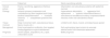
Skin damage arises from the interaction of multiple factors: trauma against the vessel due to constant pitching, compression from prolonged static postures, continuous friction of wet clothing against skin, maceration from ongoing water exposure, contact with fuel,11 and exposure to infectious agents.12 Two cutaneous syndromes associated with the journey have been described: (1) “patera foot,”12 a necrotizing infectious cellulitis due to contact with contaminated boat water that extends deeply and often requires antibiotic therapy and surgery, and (2) sterile necrotizing cellulitis,13 a sterile edema of subcutaneous tissue limited to skin and subcutis (Table 1).
Comparison of the two proposed patera travel-associated cutaneous syndromes.
| Patera foot | Sterile necrotizing cellulitis | |
|---|---|---|
| Clinical picture | Painful, necrotizing, aggressive infectious cellulitis | Inflammation and subcutaneous edema with epidermal necrosis |
| Etiology | Ischemic process (compression and cold)Infection by Gram-negative bacilli (high bacterial load)Inflammation+increased ischemia+compartment-syndrome-like process | Hypernatremic dehydration+aggressive fluid replacement+systemic inflammationEdema of subcutaneous tissueMicrocirculatory compressionSterile necrosis of skin and subcutaneous tissue |
| Tissue involved | Deep involvement (skin, subcutaneous tissue, fascia, muscle, etc.) | Limited to skin; fascia, muscle, and deep tissues spared |
| Management | Early broad-spectrum antibiotics, then targeted therapyAggressive surgical debridement | Early escharotomy-type incisions to evacuate edemaCareful fluid–electrolyte replacement |
| Prognosis | Severe (sepsis, amputations, etc.); rapid, unfavorable course | Better prognosis |
We present the epidemiologic, clinical, surgical, and outcome characteristics of 93 patients who arrived by patera to Canary shores and were admitted to our center, and we discuss the pathophysiology and management of their skin and soft-tissue lesions.
Materials and methodsWe conducted a descriptive, cross-sectional observational study including migrant patients arriving by irregular sea route who were evaluated in the Emergency Department of Complejo Hospitalario Universitario Nuestra Señora de Candelaria (CHUNSC) from January 1st through December 31st, 2023. For those admitted to the hospital, we recorded epidemiologic characteristics (age, sex, country of origin, days of travel); main admission diagnoses (metabolic acidosis, acute kidney injury, diabetes mellitus, anemia, decreased level of consciousness or shock, hypernatremic dehydration, rhabdomyolysis, skin involvement, death, and other); location and description of cutaneous lesions; surgical indication (type and number of procedures); microbiology (result of the first positive skin culture obtained within the first 4 days of admission); and admission data (length of stay, consulting services, length of ICU stay [days], and need for vasopressors). Data were retrieved from health records, laboratory reports, and microbiology reports. Data were updated through May 11th, 2024. Descriptive statistics (mean, median, standard deviation, and range for quantitative variables) were performed with IBM SPSS Statistics, version 25.0 (IBM Corp., 2017). The study met local Research Ethics Committee requirements.
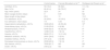
ResultsA total of 485 patients were evaluated in the Emergency Department (409 men [84.3%]), mean age 24 years (range, 0–60). A total of 93 patients (19.2%) required admission (85 men [91.4%]), mean age 23 years; mean voyage duration was 9 days (range, 6–20). Countries of origin were Senegal (35.5%), Gambia (29.0%), Mali (4.3%), Guinea-Conakry (3.2%), and Morocco (2.1%); in 25.8% origin was not recorded.
Mean inpatient length of stay was 29 days (range, 1–178). Consulting services included Internal Medicine, 61 patients (65.5%); Plastic Surgery, 32 (34.4%); ICU, 25 (26.9%); Orthopedics/Trauma, 23 (24.7%); Pediatrics, 13 (14%); Vascular Surgery, 10 (10.7%); General Surgery,7 (7.5%); Hematology, Pulmonology, and Psychiatry, 5 (5.4%) each; Nephrology, Maxillofacial Surgery, and Rehabilitation, 4 (4.3%) each; Cardiology, Dermatology, Ophthalmology, Neurology, and Obstetrics/Gynecology 3 (3.2%) each; Urology, Neurophysiology, and Pediatric Surgery, 2 (2.1%) each; and Thoracic Surgery, Neurosurgery, Gastroenterology, and Endocrinology, 1 (1.1%) each. Key clinical features are shown in Table 2. Zero patients had HIV infection; 6 had malaria; 22 (23.6%) were admitted to the ICU; 9 required vasopressors; 36 (38.7%) underwent surgery, with a mean 1.68 procedures (range, 0–4), and 5 underwent surgery for hollow-viscus perforation.
Comparison of epidemiologic, clinical, and outcome results.
| Current series | Francés Monasterio et al.13 | Rodríguez del Rosario et al.7 | |
|---|---|---|---|
| Admitted, N (%) | 93 (19.2) | 86 (ND) | 111 (12) |
| Men, N (%) | 85 (91.4) | 68 (79.07) | ND |
| Age, mean (years) | 22.9 | 24 | 25 |
| Mean voyage duration (days) | 9 | 8.91 | ND |
| Mean length of stay (days) | 29.5 | 13 | 10.2 |
| ICU admission, N (%) | 22 (23.6) | 14 (16.2) | 7 (6.31) |
| ICU admission, mean (days) | 7.6 | 5.43 | 6.5 |
| Hypernatremic dehydration, n/N (%) | 30/89 (29.6) | 46 (53.49) | 18a |
| Acute kidney injury, n/N (%) | 21/89 (23.6) | 6 (6.98) | 1a |
| Rhabdomyolysis, n/N (%) | 50/80 (62.5) | 45 (52.33) | 0a |
| Hypothermia, n/N (%) | 12/88 (13.6) | 7 (8.14) | 1a |
| Anemia, n/N (%) | 68/93 (73.1) | 10 (11.63) | 0a |
| Metabolic acidosis, n/N (%) | 24/81 (29.6) | 5 (5.81) | 0a |
| Diabetes mellitus, n/N (%) | 2/91 (2.2) | 3 (3.49) | 1a |
| Decreased consciousness or shock, n/N (%) | 20/89 (22.5) | 7 (8.14) | 0a |
| Skin lesions, n/N (%) | 76/93 (81.7) | ND | ND |
| Death, n/N (%) | 2/93 (2.2) | ND | 4a |
Quantitative variables from cited studies are shown with >1 decimal when so reported in the original publication. ND: not available.
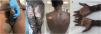
Cutaneous involvement was documented in 76 patients (81.7%). Anatomic sites were legs in 52 (68.4%), feet in 51 (67.1%), buttocks/sacrum in 43 (56.6%), arms in 29 (38.2%), hands in 27 (35.5%), genitalia in 21 (27.6%), trunk in 13 (17.1%), and head/neck in 8 (10.5%). Multiple sites were common: 14 patients (18.4%) had 1 area compromised; 15 (19.7%) had 2; 10 (13.2%) had 3; 20 (26.3%) had 4; 12 (15.8%) had 5; 5 (6.6%) had 6; none had 7 or 8. Lesions were heterogeneous and evolved over time. On the genital area and trunk, superficial erosions and small areas of skin loss were common – likely from continuous friction with wet clothing or the boat – and were generally not severe (Fig. 1). On the extremities – mainly lower limbs – early edema frequently produced blisters that progressed to necrosis and subsequent skin loss with fascial exposure (Fig. 2). Deeper or more extensive tissue loss from trauma or pressure was also seen. Lesions that initially appeared mild often evolved insidiously to involve large areas. Distal digital necrosis was frequent (Fig. 3). Patients often reported pain even when examination was unremarkable.


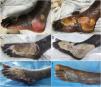
Evolution of “patera foot”-type lesions. (A and B) Arrival lesions with blisters and subsequent epidermal loss. (C and D) Established necrotic plaque and superinfected granulation tissue underneath (positive culture: Staphylococcus aureus). (E and F) Early and late postoperative appearances.

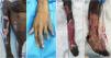
Clinical images of severe cutaneous disease associated with patera travel. (A and B) Patient with extensive involvement and distal digital necrosis of the left hand requiring amputation. (C) Patient with right hemiplegia from ischemic stroke after patera travel and extensive tissue loss of the left upper limb with mummification of the left hand, which was amputated. (D) Patient with tissue loss on the dorsum of the foot and right pretibial area with slough and hypergranulation, and digital necrosis of the 2nd–5th toes, later amputated.
Of the 76 patients with skin lesions, 31 (40.8%) required surgery for those lesions. Debridement of cutaneous necrosis was performed in 22 (71.0%); amputation in 12 (38.7%). Of the latter, 10 had limited amputations of one or more digits; 2 required more complex amputations. Coverage used split-thickness skin grafts in 21 patients (67.7%); 4 required flaps (12.9%). The remaining 45 patients (59.2%) improved with conservative care.
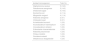
Table 3 illustrates the results of 35 positive skin cultures obtained within the first 4 inpatient days. Positive cultures were obtained in 21 of 31 surgically treated patients (67.7%), 18 of 22 debrided patients (81.8%), and 10 of 12 amputees (83.3%).
Results of the first skin culture within the first 4 inpatient days.
| Isolated microorganism | Total (%) |
|---|---|
| Staphylococcus aureus | 5 (14.3) |
| Pseudomonas aeruginosa | 5 (14.3) |
| Shewanella algae | 5 (14.3) |
| Escherichia coli | 4 (11.4) |
| Morganella morganii | 4 (11.4) |
| Klebsiella aerogenes | 2 (5.7) |
| Citrobacter koseri | 2 (5.7) |
| Acinetobacter baumanii | 1 (2.9) |
| Arcanobacterium haemolyticum | 1 (2.9) |
| Corynebacterium striatum | 1 (2.9) |
| Enterobacter cloacae/asburiae | 1 (2.9) |
| Klebsiella pneumoniae | 1 (2.9) |
| Proteus mirabilis | 1 (2.9) |
| Providencia stuartii | 1 (2.9) |
| Streptococcus disagalactiae | 1 (2.9) |
Most of the cohort originated south of Morocco, likely reflecting the more southerly departure points in current migration flows.14 This crossing is longer – both in distance and duration – and therefore migrants are more susceptible to illness. Several post-arrival clinical-worsening risk factors linked to the crossing have been identified: a death during the voyage; travel ≥4 days; >24h without water; many passengers in fair/poor condition; multiple wounds; or numerous travelers from the same boat requiring hospital referral.15
Table 2 compares our series with the only two previously published series, showing similar epidemiology and voyage duration. In our study, the mean length of stay was double that of prior reports; both the proportion of ICU admissions and mean ICU stay were also higher.
Greater clinical severity and longer admissions may relate to today's longer routes,14 more overloaded boats,5 and health care strain in a record-arrival year already impacted by the COVID-19 pandemic – factors that create bottlenecks in care delivery.
Systemic abnormalities at admission were common and generally more frequent than in prior series. Hypothermia and hypoglycemia are the two most common reasons for medical attention on arrival,9 yet we observed these were less commonly diagnosed at hospital admission7 (Table 2). Infectious findings included 6 cases of malaria and 0 HIV infections – contrasting with Francés Monasterio et al.13 who found 1 and 1, and Rodríguez del Rosario et al.7 who found 38 and 0, respectively.
Skin lesions were documented in 81.7% of charts, though milder cases likely went unnoticed and the true figure may be higher. Multiple sites – particularly lower limbs and lower back – were common, as in prior studies.12,13 An Andalusian study showed dermatologic problems were the most prevalent (24.7%), more often in lower than upper limbs and mostly superficial and mild.11 Another study,16 evaluating health problems among crew of a drifting boat after 14 days in Somalia, found 25.5% had skin involvement, mainly in lower limbs and multiple sites.
As in former studies,12,13 the need for necrosis debridement or amputation was high, often requiring subsequent coverage – mainly split-thickness skin grafts.
Multiple bacteria have been involved in the pathogenesis of skin/soft-tissue involvement in these populations,12,16–18 including Shewanella spp.,19Escherichia coli, Morganella morganii, Moraxella, Pseudomonas aeruginosa, Enterobacter, Streptococcus, Staphylococcus, and Vibrio spp. In our series, skin samples obtained within the first 4 days at the hospital (to avoid nosocomial acquisition) suggest a role for Staphylococcus aureus, Shewanella algae, E. coli, and P. aeruginosa in this syndrome and support empirical antibiotic regimens covering these organisms (Table 3).
To date, two skin/soft-tissue syndromes associated with patera travel have been described (Table 1). “Patera foot,” proposed from a 7-patient series, is an infectious cellulitis of the lower limbs – potentially aggressive and necrotizing – caused by Gram-negative bacteria (notably S. algae) in contaminated water. A compressive ischemic mechanism likely facilitates deep involvement. Low cutaneous distensibility due to edema and that typical of darker skin may aggravate the condition in a compartment-syndrome-like manner.17 Authors propose an early, aggressive antibiotic and surgical management to limit progression; amputation is sometimes required.
However, deep-tissue ischemia, distal necrosis, and amputations can occur in the absence of infection via interacting phenomena: immersion syndrome (with immobility and malnutrition as predisposing factors),20 crush injury, direct trauma, reperfusion, or hypercoagulability.21 In our cohort, 59.2% did not require surgery for resolution, and frank compartment syndrome was uncommon. Dark skin as a “risk factor”12 may be a confounder, as these populations often undertake longer routes and arrive in worse condition.
Sterile necrotizing cellulitis presents as massive, painful edema limited to skin and subcutis. Edema may result from an osmotic effect of seawater ingestion/immersion and aggressive correction of hypernatremic dehydration, collapsing cutaneous capillaries and causing secondary necrosis without extending beyond the fascia. Careful fluid–electrolyte correction and early surgical intervention with escharotomy-type incisions have been suggested to evacuate edema and halt progression.
However, in our experience, seawater ingestion is uncommon, and direct water exposure does not explain upper-limb disease. Systemic inflammation, trauma, ischemia–reperfusion, and bacterial infection/colonization – attenuated by empirical antibiotics – may play important roles in localized edema and subsequent necrosis.
Evidence for both described syndromes is limited; we likely face a broader patera-associated skin and soft-tissue syndrome (Fig. 4) – a spectrum in which advanced “patera foot” represents the severe end, whereas milder forms respond to antibiotics or are self-limited.

Proposed pathophysiology of patera-associated skin and soft-tissue syndrome. Non-freezing cold injury (immersion/trench foot) from prolonged exposure to 0–15°C and humidity causes neurovascular damage and sustained vasoconstriction; fatigue, malnutrition, dehydration, immobility, and Afro-Caribbean ancestry are predisposing factors. Crush injury leads to soft-tissue ischemia, direct tissue damage, and impaired venous return. Subsequent reperfusion of previously hypoperfused areas triggers soft-tissue edema and systemic release of damage-associated molecules, worsening the condition. Trauma from wave motion and friction with clothing/boat causes cutaneous injury, exacerbated by cold-related sensory alteration and peripheral neuropathies. Compression of the common peroneal nerve – typically from prolonged squatting or external compression – is another factor, partially explaining pretibial and dorsal-foot lesions. Boat bilge water contains stagnant salt water, organic waste, chemical irritants, and high bacterial loads, favoring colonization/infection. Other contributors include malnutrition/dehydration, systemic inflammation, and hypercoagulability.
When skin/soft-tissue infection is suspected, early management is essential: appropriate comorbidity control, systematic cultures, and antibiotic coverage (Fig. 5). Broad-spectrum empiric therapy should cover S. aureus and Gram-negative bacteria including Pseudomonas; because of the marine setting, coverage for Shewanella spp. and Vibrio spp. should be considered,22 even though Vibrio was not identified in our series.

Proposed hospital management algorithm for skin and soft-tissue lesions after patera arrival. *In hospitalized patients, consider coverage for Staphylococcus aureus, Gram-negative bacteria (including Pseudomonas aeruginosa and Shewanella), and Vibrio spp.; broad-spectrum regimens with antipseudomonal agents plus addition of doxycycline are reasonable. **The role of escharotomy-type incisions has been proposed by some authors13 to slow progression of cutaneous/subcutaneous edema without infection criteria; however, its utility has not been demonstrated.
Special attention should be paid to pain frequently reported by patients without significant visible lesions. This may reflect contusions or incipient necrosis, potentially obscured by the challenges of examining darkly pigmented skin. The role of escharotomy-type incisions is controversial and warrants future study.
One study assessed hospital workload by service,7 with results similar to ours: among 111 discharges, 65 were from Internal Medicine, 16 from Trauma, and 7 from Intensive Care – only 1 from Plastic Surgery. Close collaboration across services is essential, given the cross-cutting nature of these clinical presentations (Table 2).
LimitationsKey limitations include the retrospective design; challenges caring for this population (language barriers and skin phototype); altered clinical presentations after empiric treatments started immediately on landfall; lack of standardized care pathways for occasionally massive contingents; and underestimation of systemic and dermatologic conditions not requiring admission and thus not captured in this study.
ConclusionsClinical heterogeneity and limited knowledge of optimal management contribute to prolonged admissions and occasional severe outcomes. Most patients are young, otherwise healthy men who on arrival present with anemia, dehydration, rhabdomyolysis, renal failure, and complex skin lesions that often drive prognosis.
Conflicts of interestNone declared.


