


Mohs micrographic surgery (MMS) is known to be a time- and resource-consuming procedure; therefore, methods to optimize MMS workflow are always welcome.
In this brief communication we wanted to share our way to perform MMS processing using tap water (TW) embedding and methylene blue (M-blue) stain. As far as we know, both TW embedding and M-blue stain have never been reported for MMS processing. However, they have been historically used for the intraoperative pathologic evaluation of other surgical specimens.1,2 Therefore, the application of this processing method to MMS is a “back to basics” of intraoperative pathology. Feasibility of this processing is supported by the data of 261 consecutive MMS cases collected in a prospective database from February 2022 through August 2024.
Standard optimal cutting temperature (OCT) embedding involves using embedding devices, embedding molds, or glass slides and a spirit lever or a heat extractor. The reason to use OCT is to quickly reach an optimal cutting temperature to avoid well-known frozen artifacts. However, in our experience TW does not generate more frozen artifacts than OCT with faster freezing times.
Our MMS processing follows these steps: (1) after surgical excisions, MMS specimens are processed as per standard MMS technique; (2) a cryostat tissue holder (chuck) is prepared in the cryostat with a layer of frozen TW (i.e., an ice bed) to have a smooth surface to attach and lay the sections flat; (3) the chuck is taken out from the cryostat and the MMS section is attached to it; (4) drops of TW are added around the section to complete the embedding; (5) the chuck is, then, put back in the cryostat and the heat extractor is applied; (6) after 10–15s, the section is ready to be trimmed and cut. Trimming is usually reduced because there is no layer of OCT or other embedding medium to remove. The Mohs technician places progressive layers on a glass slide, making sure to include the entire section. Finally, slides are stained with M-blue with the following ultra-rapid protocol: 5s immersion in M-blue (Kühne's Methylene Blue Phenicated solution, QCA, Amposta, ES) and 5s wash in TW. Slides are then dried with paper and passed to the pathologist and Mohs surgeon for evaluation. To further speed up the processing, no cover slip is applied, and no mounting media is needed. They will be applied later when the slides are ready to be scanned (Aperio GT450, Leica, Wetzlar, DE). The steps of this MMS processing and some performance tips are showns in video 1. Quality of the slides obtained with our method is shown in Figs. 1 and 2 and video 1.
 of a nodular basal cell carcinoma in this final layer of a 2-section Mohs micrographic surgery specimen (263nm/pixel, 1.08×). Slide quality is good and adipose tissue is present.")
 adipose rich Mohs micrographic surgery specimen (263nm/pixel, 0.52×). At 5× the excellent frozen fat quality is evident.")
The use of TW embedding can be a reason form concern in some readers. Such concern also worried the main author (R.M.) when he started performing MMS after his fellowship. However, after more than 2 years, we agree that using TW embedding does not negatively affect the quality of the slides. Indeed, it seems to be very helpful for cutting through adipose tissue, a known problem of MMS.3 Therefore, TW embedding maintains frozen section skin quality, while enhancing fat quality without the need for further freezing with ice spray or liquid nitrogen (Figs. 1 and 2). Moreover, it reduces trimming time because no layer of embedding medium covers the specimen surface. Finally, using TW instead of OCT lowers the costs of MMS processing.
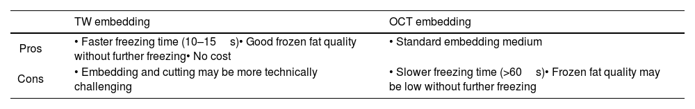
One drawback of TW embedding may be the difficulty to keep the MMS section flattened once flipped over on the chuck, because it will not be “floating” in the OCT. Indeed, the skin edge may drop below the horizontal plane of the section, especially if cutting a single-section MMS. Nevertheless, the skin edges can be elevated (video 1). Comparison between TW and OCT embedding is shown in Table 1.
Comparison between tap water (TW) and optimal cutting temperature (OCT) embedding.
| TW embedding | OCT embedding | |
|---|---|---|
| Pros | • Faster freezing time (10–15s)• Good frozen fat quality without further freezing• No cost | • Standard embedding medium |
| Cons | • Embedding and cutting may be more technically challenging | • Slower freezing time (>60s)• Frozen fat quality may be low without further freezing |
The use of M-blue stain may be less surprising. M-blue stain is practically identical to toluidine blue (T-blue) stain. T-blue is very well known in literature for enhancing visualization of skin cancers normally treated by MMS. This enhancement is due to a phenomenon called metachromasia, whereby the T-blue staining of the mucopolysaccharide stroma around tumor cells shifts towards pink or magenta.4 Although M-blue shows the same metachromatic phenomenon as T-blue (Fig. 1 and video 1), but it has never been reported as a stain of MMS layers. There are not clear advantages or drawbacks associated with the use M-blue instead of T-blue; it is just a matter of preference, cost, and availability. Remarkably, our 10s M-blue protocol is way faster than previously described T-blue protocols.4,5
Although there have not been recurrences of skin cancers treated with this MMS processing, follow-up time – 2.5 years – is very short to draw any conclusions on recurrence rates.
In conclusion, we reported how we do the MMS processing illustrating two novel features: TW embedding and ultra-rapid M-blue stain. These new features speed up MMS processing without seemingly affecting slides quality and clinical results. Review of the slides without mounting media or cover slips further speeds up the process, which is especially important when multiple sections are required. Longer follow-up and comparative studies are needed to confirm our experience.
Conflict of interestThe authors declare that they have no conflict of interest.


