Reverse isotopic phenomenon, or isotopic non-response, is an extremely rare phenomenon characterised by the absence of a skin disease at the site of another unrelated skin disease which is already healed.1 Sparing occurs at the healed site of an inflammatory disease. Most reports of reverse isotopic phenomenon show the sparing of the skin previously involved in herpes zoster.2
We describe a case of Drug Reaction with Eosinophilia and Systemic Symptoms (DRESS) syndrome due to carbamazepine with isotopic non-response at the site of healed herpes zoster.
A 60 year male presented to us with complaints of fever, breathlessness and red itchy rash over body since 7 days. The patient had developed herpes zoster on the lower part of the right chest around two months back, and was prescribed Tab Carbamazepine by a local physician for the residual pain.
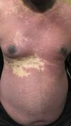
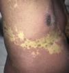
On examination, the patient was found to have generalised swelling, particularly prominent over the periocular areas. He was febrile (100.6 F) and had tachycardia (heart rate-110/min) and tachypnea (respiratory rate-22/min). The abdomen was distended and bowels sounds were not heard. There was mildly tender lymphadenopathy in inguinal and axillary areas. Cutaneous examination revealed erythematous maculopapular lesions predominantly distributed over the face, trunk and proximal extremities with tendency to coalesce such that the whole of the trunk was involved except the right T9 dermatome which was characteristically spared. [Fig. 1] The skin of the right T9 dermatome showed hyperpigmented scars of the healed herpes zoster. [Fig. 2] The rest of the cutaneous and systemic examination was normal.
Investigations revealed leucocytosis (TLC- 15500/ cu mm; normal- 4000-11000/ cu mm) with eosinophilia (AEC- 1580/ cu mm; normal- 40-440/ cu mm). There was elevation of liver enzymes (SGOT- 660 IU; normal- 7-40 IU, SGPT- 590 IU; normal- 7-56 IU) and increased blood urea (120mg/dl; normal- 10-40mg/dl) and serum creatinine (1.9mg/dl; normal- 0.3-1.2mg/dl). The patient had a normal chest radiograph and a gas filled distended bowel suggestive of paralytic ileus. The patient had hyperamylesemia (260 U/l; normal- 40-140 U/l) suggestive of pancreatitis. He subsequently developed fatal acute respiratory distress syndrome. A diagnosis of DRESS syndrome was made based on the RegiSCAR criteria.3
DRESS syndrome is a rare idiosyncratic reaction reaction to a drug characterised by skin rash, fever, lymphadenopathy, eosinophilia and multiorgan involvement. It is commonly caused by anticonvulsant medications, allopurinol, minocycline and antiretroviral agents like abacavir. Pathogenesis is unclear but it is believed to occur in patients who metabolise the drugs and its active metabolites slowly. A genetic predisposition exists and Human Herpesviruses has been associated. The cross reaction of antiviral T cells with the culprit drug has been proposed to mediate this syndrome.4 However, the sparing of the healed herpes zoster site casts doubt regarding this theory.
The possible reason for the sparing of a skin lesion to a secondary insult is unclear. The rash of DRESS may result from alteration of the local immune response by a virus itself or by production of Th1 cytokines like TNF-alpha, IFN-gamma, IL-2 and IL-5.2 The loss or hypoactivity of Langerhans cells at the site of herpes zoster has been demonstrated and may play a role in this sparing phenomenon.5 Also, herpesvirus infected keratinocytes have decreased expression of MHC and ICAM-1, hindering their function as antigen presenting cells and inhibiting T cell response, thereby leading to the skin rash of DRESS.6
Isotopic non-response needs to be differentiated from isomorphic non-response or Renbök phenomenon. The site of another unrelated skin disease is spared in both of the above phenomenon. This disease is still active in Renbök phenomenon while it is healed in the isotopic non-response.1
Very few cases of reverse isotopic phenomenon have been reported in literature. There reports have shown the sparing of the sites of vaccination,7 herpes zoster2,5,6,8,9 and burn scar10 that have occurred in diverse conditions like epidermal necrolysis,2 erythema multiformae,8 cutaneous lymphomas,5 leprosy9 and annular elastolytic giant cell granuloma.10 [Table 1] Ours is probably the first case of reverse isotopic phenomenon in DRESS syndrome.
Details of cases of isotopic non-response reported in literature.
| N.° de caso | Estudio | Edad/sexo | Enfermedad secundaria | Sitio de la preservación | Enfermedad primaria |
|---|---|---|---|---|---|
| 1 | Nuestro caso | 60 años/varón | Síndrome DRESS | Tronco (dermatoma T9 derecho) | Herpes zóster |
| 2 | Tenea D2 (2010) | 39 años/mujer | Síndrome Stevens-Johnson | Lado derecho del tronco | Herpes zóster |
| 3 | Kannangara AP et al.6 (2008) | 53 años/mujer | Síndrome sobreposición SJS-TEN | Nervio craneal V1 izquierda | Herpes zóster |
| 4 | Kannangara AP et al.6 (2008) | 62 años/varón | Eritrodermia | C3-C4 izquierda | Herpes zóster |
| 5 | Park H et al.7 (2008) | 67 años/mujer | Eritema multiforme | Tronco (dermatoma T3) | Herpes zóster |
| 6 | Twersky JM et al.5 (2004) | 58 años/varón | Linfoma cutáneo células T | Tronco (dermatoma T8 izquierdo) | Herpes zóster |
| 7 | Jain R et al.8 (2003) | ND | Lepra «borderline» | ND | Herpes zóster |
| 8 | Jain R et al.8 (2003) | ND | Lepra «borderline» | ND | Herpes zóster |
| 9 | Okaya-Bayazit E et al.9 (1999) | 72 años/mujer | Granuloma elastolítico anular de células gigantes | Antebrazo izquierdo | Cicatriz quemadura |
| 10 | Nasca MR et al.10 (1995) | 53 años/varón | Acné esteroideo | Espalda | Sitio irradiación Rx |
| 11 | Huilgol SC et al.11 (1995) | 74 años/varón | Granuloma anulargeneralizado | Brazo izquierdo | Punto de vacunación |
DRESS: drug reaction with eosinophilia and systemic symptoms; ND: no disponible; Rx: rayos X; SJS-TEN: Stevens-Johnson syndrome-toxic epidermal necrolysis.
The authors declare that they have no conflicts of interest.
Please cite this article as: Adil M, Amin SS, Dinesh Raj R, Tabassum H. Reverse Isotopic Phenomenon in Drug Reaction with Eosinophilia and Systemic Symptoms. Actas Dermosifiliogr. 2019;110:613–615.


