


The gut microbiota interacts with the immune system and plays an important role in many inflammatory diseases such as psoriasis, although the exact mechanisms in this disease are not yet well understood.
ObjectivesTo characterize differences in the microbiota between patients with psoriasis and healthy controls, and to assess the relationship between these differences and the interleukins involved in psoriasis.
MethodsA cross-sectional observational study was conducted in which sociodemographic data, blood samples, and stool samples were collected from patients with psoriasis and healthy controls attending our center between June 2019 and May 2020. Cytokines (interleukin (IL) 17, 22, 23, 31, 33, 36, interferon (IFN) γ, and transforming growth factor (TGF) β) were analyzed using ELISA, and microbiota was analyzed through 16S amplicon sequencing.
ResultsThirty-six patients and 23 controls were included. Absolute abundance analysis found a higher abundance of the phylum Synergistota in the control group (p<0.05). Differential abundance analysis found higher abundance of the genus Subdoligranulum and Lactobacillus, and the species Bacteroides plebeius (p<0.05), and lower abundance of the species Senegalimassilia anaerobia and the genus Ruminococcus (p<0.05) in the psoriasis group. A relationship was observed between Subdoligranulum and TNFα, IL17, IL22, IL23, IL31, IL33, IL36, IFNγ, and TGFβ (p<0.05), as well as between Lactobacillus and IL17, IL23, IL36, TNFα, and TGFβ (p<0.05).
ConclusionsSignificant alterations in the gut microbiota of patients with psoriasis were detected and a relationship with inflammatory interleukins, suggesting their involvement in the disease. These findings could aid in the development of future probiotic treatments for psoriasis.

Psoriasis involves a complex interaction of factors. In its pathogenesis, dysregulation of the interleukin (IL)-23–Th17–IL-17 axis plays a crucial role and leads to elevated levels of tumor necrosis factor (TNF)-α, IL-23, and IL-17. Additionally, increased levels of interferon (IFN)-α, IFN-γ, IL-2, IL-6, IL-8, IL-12, IL-15, and IL-18 contribute to the chronic inflammatory state observed in psoriatic lesions. Various factors have been implicated, including genetics and environmental triggers, and recent research highlights the potential role of the microbiota, suggesting that alterations in the cutaneous and gut microbiota may influence disease pathogenesis and progression.1
The concept of the microbiota was described in 2001 as “the ecological community of commensal, symbiotic, and pathogenic microorganisms that share our body space.” It refers to the population of bacteria, fungi, and viruses inhabiting our skin, gut, and other sites.2 This complex community plays a crucial role in maintaining health and homeostasis, and changes in its composition – known as dysbiosis – have been linked to inflammatory diseases such as acne and atopic dermatitis.3
In psoriasis, this relationship has not been studied as extensively. Existing studies are limited, often involving small patient cohorts and lacking rigorous control groups.4–7 This gap underscores the need for more comprehensive studies to elucidate the role of the gut microbiota in psoriasis.
A study was conducted comparing the gut microbiota of patients with psoriasis with that of healthy controls, taking into consideration clinical characteristics of psoriasis and a detailed analysis of inflammatory cytokine levels. By correlating these clinical parameters with microbiota profiles, we aim to provide deeper insight into the potential role of gut dysbiosis in the pathogenesis of psoriasis.
Material and methodsStudy design and patientsWe conducted a cross-sectional study including adult patients with psoriasis seen in our department. Exclusion criteria were the use of antibiotics within the previous 3 months, consumption of probiotics or adherence to a specific diet within the last 6 months, as well as the presence of cirrhosis, neoplasms, inflammatory bowel disease, or other autoimmune diseases. Healthy controls met the same criteria.
After informed consent was obtained, clinical data were collected along with stool samples for gut microbiota analysis and blood samples for standard biochemical tests, complete blood counts, and interleukin measurements.
Interleukin measurementSerum enzyme-linked immunosorbent assays (ELISA) were performed for TNF-α, IL-17, IL-22, IL-23, IL-31, IL-33, IL-36, IFN-γ, and TGF-β (Invitrogen™ kits, Thermo Fisher Scientific, Waltham, MA, USA). Samples were analyzed in triplicate and read using a Sunrise microplate reader (Tecan, Männedorf, Switzerland). The lower limit of detection was 5pg/mL. Standard curves were generated for each plate, and the optical densities of the zero standard were subtracted from all other standards, controls, and samples to obtain corrected concentrations.
Sample preparation and sequencingGenomic DNA was extracted from stool samples using the QIAamp PowerFecal DNA Kit (Qiagen, Hilden, Germany), and gut microbiota composition was analyzed by sequencing the hypervariable V3–V4 region of the 16S rRNA gene on an Illumina MiSeq platform.
Quality control and data processingA mean 141,200 paired Illumina reads per sample was obtained. Quality control was performed using fastqc8 and MultiQC.9 The DDAA2 pipeline (v1.16.0) was then used to generate the amplicon sequence variant (ASV) table from raw reads. Duplicates, noise, and chimeras were removed using default settings. Taxonomic annotation of ASVs was performed using the SILVA database (v138.1).10 A rarefaction curve was calculated using the R package vegan and inspected to assess taxon-count saturation. A phylogenetic tree of ASVs was constructed using Phangorn, and this tree was used to compute UniFrac and weighted UniFrac distances.11 The ASV count matrix was rarefied to the minimum read count per sample.
Alpha diversity analysisAlpha-diversity measures capture taxon diversity within each sample. Using the phyloseq package (v1.44.0),12 observed richness and Chao1 were calculated, as well as Shannon, inverse Simpson, and Fisher's alpha diversity indices. To test statistically significant differences across samples from controls and patients with psoriasis, the Wilcoxon rank-sum univariate test was first applied. To control for potential confounders, linear regression models including psoriasis and one or more covariates (body mass index [BMI], sex, age, smoking, alcohol intake, and number of reads) were used.
Beta diversity analysisBeta diversity refers to differences in community composition between samples. The phyloseq package (v1.44.0)12 was used to compute weighted UniFrac distances11 and to ordinate data points in a lower-dimensional space using principal coordinates analysis (PCoA). To evaluate shifts in the overall distribution of ASV abundance, PERMANOVA was applied using the adonis2 function of the vegan package, based on weighted UniFrac distances. Separate tests were run for each variable (psoriasis, BMI, sex, age, smoking, alcohol intake, and number of reads), followed by an additional test combining confounders identified as significant in individual models.
Taxonomic profilingTo display taxonomic composition at the phylum level, the plot_bar function of phyloseq was used. ASV counts belonging to the same genus were then aggregated, and the 15 genera with the highest mean abundance were plotted using the fantaxtic R package.
Differential abundance analysisTo test for taxa with differential abundance between conditions, ASVs with <1 count in at least 3 samples (prevalence <6.25%) were removed, and DESeq2 with the Wald test was applied. Furthermore, DESeq2 models that included psoriasis status plus covariates (sex, BMI, age, smoking, or number of reads) were also fitted. The likelihood ratio test (LRT) was used to compare these models with models that included only confounding variables. Due to limited statistical power, only a few ASVs had a p-value <0.05 and none had an adjusted p-value <0.05.
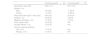
ResultsClinical and demographic characteristicsA total of 36 patients with psoriasis and 24 controls were included (Table 1). The mean age in the psoriasis group was 46.6 years (SD, 11.7), higher than in the control group (40.7 years, SD, 17.0). Sex distribution was similar across groups (52.8% men in the psoriasis group vs 45.8% in the control group). The mean BMI was comparable between groups (26.6±5.5 and 26.3±5.4kg/m2, mean±SD for psoriasis and control groups, respectively). The prevalence of smokers was significantly higher in the psoriasis group (55.6% vs 20.8% in controls). In addition, the incidence rate of metabolic syndrome was higher in patients with psoriasis (30.6% vs 12.5% in controls). The median Psoriasis Area and Severity Index (PASI) for the psoriasis group was 3.6 (IQR, 4.35). Similarly, 27.8% of the psoriasis group exhibited psoriatic arthritis (PsA), and 94.4% reported prior systemic treatment, 47.2% oral and 47.2% biologic.
Characteristics of patients with psoriasis and control group.
| Psoriasis group(N=36) | Control group(N=24) | |
|---|---|---|
| Age (years), mean (SD) | 46.6 (11.7) | 40.7 (17.0) |
| Gender, n (%) | ||
| Male | 19 (52.8) | 11 (45.8) |
| Female | 17 (47.2) | 13 (54.2) |
| Body mass index (kg/m2, mean (SD)) | 26.6 (5.5) | 26.3 (5.4) |
| Smoker, n (%) | 20 (55.6) | 5 (20.8) |
| Metabolic syndrome, n (%) | 11 (30.6) | 3 (12.5) |
| PASI, median (IQR) | 3.6 (4.35) | N/A |
| Psoriatic arthritis, n (%) | 10 (27.8) | N/A |
| Previous systemic treatment | ||
| Oral, n (%) | 17 (47.2) | N/A |
| Biologic, n (%) | 17 (47.2) | N/A |
IQR: interquartile range; N/A: not applicable; SD: standard deviation.
Observed richness and Chao1 were computed, as well as Shannon, inverse Simpson, and Fisher's alpha diversity indices. No significant differences were found for any diversity index between groups using the Wilcoxon test (Fig. 1A). No significant differences were found when using linear regression models including confounding variables (sex, smoking, alcohol intake, BMI, and age). Among these variables, only BMI showed a marginal yet significant association with observed richness and Chao1 (R2=0.07, p=0.04) (Supplementary Fig. 1). This suggests that microbial diversity within individuals was similar regardless of whether they had psoriasis. No differences in alpha diversity were found between patients with and without PsA (Fig. 1B).
 Alpha diversity according to psoriasis vs healthy controls. (B) Alpha diversity according to the presence of psoriatic arthritis.")
The PCoA of weighted UniFrac distances showed that samples did not cluster according to psoriasis diagnosis (Fig. 2A), nor, among patients, according to the presence of PsA (Fig. 2B). Consistently, the PERMANOVA was not significant, and cutaneous psoriasis explained 1.9% of the variation in microbiota composition (Fig. 2A). Sex and smoking also did not explain a significant portion of variation in microbiota composition (1.4% and 2.5%, respectively; p>0.05) (Supplementary Figs. 2A and B). BMI, on the other hand, explained 4% of the variation in microbiota composition (p<0.05). A BMI gradient can be observed along the 2nd axis of the PCoA, with high-BMI individuals clustering at the bottom of the plot and low-BMI individuals at the top (Supplementary Fig. 2C). Age explained 4.9% of the overall variation in composition (p<0.05) (Supplementary Fig. 2D).
. (A) Beta diversity according to the presence (red) or absence (blue) of psoriasis. No clustering according to psoriasis diagnosis is observed. (B) Beta diversity according to the presence of psoriatic arthritis. No segregation of cases is observed according to this variable.")
Beta diversity based on principal coordinates analysis (PCoA). (A) Beta diversity according to the presence (red) or absence (blue) of psoriasis. No clustering according to psoriasis diagnosis is observed. (B) Beta diversity according to the presence of psoriatic arthritis. No segregation of cases is observed according to this variable.
Within patients with psoriasis, the relationship between alpha-diversity indices and BMI did not persist, likely due to sample size limitations, but the relationships with beta diversity and BMI and age did persist, explaining 5% and 4.8% of variation, respectively (PERMANOVA, p<0.05).
Furthermore, the PASI score showed a significant relationship with beta diversity, explaining 5% of the variation within the psoriasis group, although it was not associated with any alpha-diversity index. The presence of a family history did not show significant relationships with either alpha or beta diversity (Supplementary Fig. 3). Finally, prior treatments, whether classic systemic or biologic, did not appear to be associated with either alpha or beta diversity (Supplementary Fig. 4).
Taxonomic analysisAnalysis of absolute abundances revealed differences in the phylum Synergistota, which was less represented in patients with psoriasis (p<0.05) (Fig. 3A), although the total abundance of this phylum was low in both groups. A summary of species found in both groups is shown in Fig. 3B.
 Total abundance by phylum. This panel shows total phylum-level abundance in both groups: psoriasis and control. The phylum Synergistota appears less abundant in psoriasis, although overall abundance is low in both groups. (B) Relative abundance by species. This panel displays the relative abundance of the different species in both groups: control (top) and psoriasis (bottom). (C) Volcano plot for differential abundance analysis. This plot shows the results of the differential abundance analysis. ASVs with no differences across groups are shown in green. ASVs with differential abundance in psoriasis are shown in red – lower abundance on the left and higher abundance on the right.")
Total abundance by phylum, relative abundance by species, and volcano plot for differential abundance analysis. (A) Total abundance by phylum. This panel shows total phylum-level abundance in both groups: psoriasis and control. The phylum Synergistota appears less abundant in psoriasis, although overall abundance is low in both groups. (B) Relative abundance by species. This panel displays the relative abundance of the different species in both groups: control (top) and psoriasis (bottom). (C) Volcano plot for differential abundance analysis. This plot shows the results of the differential abundance analysis. ASVs with no differences across groups are shown in green. ASVs with differential abundance in psoriasis are shown in red – lower abundance on the left and higher abundance on the right.
The differential abundance analysis (DAA) identified increased levels of Subdoligranulum sp. (ASV188), Lactobacillus sp. (ASV207), and Bacteroides plebeius (ASV45), and decreased levels of Senegalimassilia anaerobia (ASV225) and Ruminococcus sp. (ASV250) in patients with psoriasis vs controls. After adjusting for confounding factors including BMI, age, sex, and smoking, only Subdoligranulum sp. remained significant (Fig. 3C), indicating a strong positive association between the abundance of this genus and psoriasis.
Additionally, a DAA was conducted considering only patients with psoriasis. No differences in any ASV were identified according to PsA status, family history, PASI, or prior systemic/biologic treatments, likely due to sample size limitations.
Relationship between interleukins and microbiotaMeasurement of inflammatory interleukins showed significantly elevated levels of all measured cytokines in patients with psoriasis (Fig. 4A) (p<0.05). When assessing the relationship between ASVs and inflammatory interleukins, only a few differentially abundant ASVs showed group-specific correlations with certain cytokines (Fig. 4B).
 Interleukin levels in peripheral blood samples. This panel shows mean interleukin levels with standard deviation for both groups: control (blue) and psoriasis (red). All interleukins show statistically significant differences. (B) Heatmap of the correlation between species abundance and interleukin levels. This heatmap represents the correlation between ASV abundance and interleukin levels. The top bar represents the p-value for each correlation (gray for p<0.05 and black for adjusted p<0.05). The 2nd bar indicates whether ASVs are more (red) or less abundant (blue) in psoriasis. The top rectangle includes all samples; the one in the middle represents the control group; the bottom one represents the psoriasis group. Colors in each square reflect whether interleukin levels are higher (red) or lower (blue). Red squares for more abundant ASVs represent positive correlations; blue squares represent negative correlations. For less abundant ASVs, blue squares indicate positive correlations and red squares indicate negative correlations.")
Interleukin levels in peripheral blood samples and heatmap of correlations between species abundance and interleukin levels. (A) Interleukin levels in peripheral blood samples. This panel shows mean interleukin levels with standard deviation for both groups: control (blue) and psoriasis (red). All interleukins show statistically significant differences. (B) Heatmap of the correlation between species abundance and interleukin levels. This heatmap represents the correlation between ASV abundance and interleukin levels. The top bar represents the p-value for each correlation (gray for p<0.05 and black for adjusted p<0.05). The 2nd bar indicates whether ASVs are more (red) or less abundant (blue) in psoriasis. The top rectangle includes all samples; the one in the middle represents the control group; the bottom one represents the psoriasis group. Colors in each square reflect whether interleukin levels are higher (red) or lower (blue). Red squares for more abundant ASVs represent positive correlations; blue squares represent negative correlations. For less abundant ASVs, blue squares indicate positive correlations and red squares indicate negative correlations.
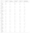
Focusing on the 5 ASVs with significantly different abundances between groups (Subdoligranulum sp., Lactobacillus sp., B. plebeius, S. anaerobia, and Ruminococcus sp.), ASVs with greater abundance in psoriasis showed positive correlations with cytokines, whereas those with lower abundance showed negative correlations (Table 2). Specifically, a significant positive correlation was found between Subdoligranulum sp. and all cytokines (TNF-α, IL-17, IL-22, IL-23, IL-31, IL-33, IL-36, IFN-γ, and TGF-β) (p<0.05), as well as between Lactobacillus sp. and IL-17, IL-23, IL-36, TNF-α, and TGF-β (p<0.05). Moreover, a significant correlation was found between B. plebeius and IL-22 (p<0.05).
Correlation between species and phyla with differential abundance in psoriasis and controls, and interleukins.a
| Interleukin | Lactobacillus sp.(ASV207) | Bacteroides plebeius(ASV45) | Subdoligranulum sp.(ASV188) | Ruminococcus sp.(ASV250) | Senegalimassilia anaerobia(ASV225) |
|---|---|---|---|---|---|
| IL-17 | |||||
| Pearson correlation | 0.29* | 0.16 | 0.46* | −0.15 | 0.12 |
| Spearman correlation | 0.35* | 0.23 | 0.44* | 0.02 | 0.15 |
| IL-22 | |||||
| Pearson correlation | −0.07 | 0.01 | 0.18 | −0.35* | −0.27 |
| Spearman correlation | 0.18 | 0.92 | 0.29* | −0.11 | −0.06 |
| IL-23 | |||||
| Pearson correlation | 0.34* | 0.26 | 0.45* | −0.22 | −0.06 |
| Spearman correlation | 0.38* | 0.28* | 0.41* | −0.04 | 0.04 |
| IL-31 | |||||
| Pearson correlation | 0.28* | 0.14 | 0.43* | −0.19 | −0.06 |
| Spearman correlation | 0.36 | 0.32* | 0.50* | 0.04 | 0.10 |
| IL-33 | |||||
| Pearson correlation | 0.24 | 0.17 | 0.41* | −0.18 | 0.01 |
| Spearman correlation | 0.32* | 0.27 | 0.46* | −0.06 | 0.04 |
| IL-36 | |||||
| Pearson correlation | 0.33* | 0.20 | 0.42* | −0.22 | −0.07 |
| Spearman correlation | 0.33* | 0.25 | 0.42* | −0.05 | 0.04 |
| TNF-α | |||||
| Pearson correlation | 0.30* | 0.24 | 0.44* | −0.26 | −0.16 |
| Spearman correlation | 0.32* | 0.30* | 0.44* | −0.04 | −0.05 |
| IFN-γ | |||||
| Pearson correlation | 0.23 | 0.15 | 0.36* | −0.22 | −0.15 |
| Spearman correlation | 0.32* | 0.25 | 0.40* | −0.01 | 0.03 |
| TGF-β | |||||
| Pearson correlation | 0.33* | 0.20 | 0.41* | −0.31 | −0.17 |
| Spearman correlation | 0.37* | 0.29* | 0.44* | −0.08 | 0.01 |
IL: interleukin; TNF: tumor necrosis factor; IFN: interferon; TGF: transforming growth factor; ASV: amplicon sequence variant.
This table shows the correlation between the 5 species and phyla that exhibited differential abundance between both groups and the levels of inflammatory interleukins. Pearson and Spearman correlation coefficients are shown. Statistically significant values (p<0.05) are marked with an asterisk (*).
Among ASVs with reduced abundance, a significant negative correlation was found between Ruminococcus sp. and IL-22 and TGF-β (p<0.05), and between S. anaerobia and elevated IL-17 levels in healthy controls only (p<0.05).
DiscussionThe relationship between dysbiosis and increased psoriasis risk is a topic of growing interest. Most available studies on gut microbiota in patients with psoriasis report an increase in the Firmicutes/Bacteroidetes (F/B) ratio in the gut microbiota of these patients.6,7,13,14 This conclusion is contradicted, however, by another study reporting an alteration of the F/B ratio favoring the phylum Bacteroidetes.6 In our study, no significant differences were found in the phyla Firmicutes or Bacteroidetes. The article by Chen et al. shows that the gut microbiota of patients with psoriasis is significantly different from that of controls only in the group with BMI <25, and that the gut microbiota of these patients differs from that of controls regardless of treatment status, suggesting persistence of microbiota alterations even after treating psoriasis.7 Furthermore, other studies report decreased richness and diversity indices.4,5,15 In our case, such differences were not found. Of note, the wide variability in results among these studies, which some authors attribute to differences in disease severity or comorbidities such as obesity.5,6,13 Another additional reason for this variability is dietary differences among patients.14 Studies involving patients from the same region and socioeconomic status, or controlling for diet, should yield more consistent results.
Regarding our objective of determining differences between patients with psoriasis and healthy controls, although no differences were found in alpha or beta diversity, significant differences were identified in taxonomic analyses and, notably, in the relationship with pro-inflammatory ILs. The decrease in abundance of the phylum Synergistota, as observed in patients with psoriasis in our study, has been associated in the literature with reduced risk of Parkinson disease and increased risk of Alzheimer disease, diabetic retinopathy, and ankylosing spondylitis.16
Among the genera and species that showed differences in the DAA, the genus Subdoligranulum sp. – associated in our study with increased levels of all pro-inflammatory ILs – has been linked to inflammatory diseases such as juvenile idiopathic arthritis, rheumatoid arthritis, Sjögren syndrome, and inflammatory bowel disease.17
Our findings are consistent with the literature, which has reported increased abundance of B. plebeius18 and decreased abundance of Ruminococcus sp.19 in patients with psoriasis. Elevated abundance of B. plebeius has been associated with biologic therapy in patients with psoriasis (which is particularly relevant given that nearly half of the patients in our study had received biologic therapy) and with mood disorders. This is noteworthy, as the prevalence of depression is much higher in patients with psoriasis than in the healthy population. The reduced abundance of Ruminococcus sp. – which in our analysis was associated with higher levels of inflammatory ILs – has previously been linked to psoriatic disease, but also to elevated C-reactive protein and erythrocyte sedimentation rate,20 ankylosing spondylitis, and coronary artery disease. Decreased abundance of S. anaerobia has not previously been associated with psoriasis,21 and in our analyses it showed an association with increased IL-17 levels in healthy controls.
The association between psoriasis and increased abundance of Lactobacillus sp., along with higher levels of inflammatory ILs, remains unclear, especially considering that many probiotics commercially available contain species from this genus.
Finally, among patients with psoriasis, we found a series of factors with higher prevalence – including smoking and metabolic syndrome, which also encompasses obesity – that have been associated with gut microbiota alterations and could act as confounders in the study.22 Another potential confounder is the high percentage of patients with psoriasis undergoing active treatment with oral and/or biologic agents, which, although supported by limited evidence, have been described as potential modulators of gut microbiota.18 The low psoriasis severity of the patients included, as measured by the PASI score, may also influence the results.
This study has certain limitations. One potential limitation is the use of 16S sequencing for ASV identification. This technique does not allow bacterial identification at the species level nor does it provide functional information. This is noteworthy because, for instance, within the genus Bacteroides, some species have anti-inflammatory properties while others may be pro-inflammatory; 16S sequencing does not allow discrimination between them. For future studies, we propose the use of whole metagenomic sequencing, which would provide greater precision and functional insight into microbiota characterization. Another limitation is the sample size, which may limit external validity. Third, the single-center design may restrict the applicability of findings to other regions. Additionally, the study design only allows observation of associations, leaving a gap in understanding the underlying pathophysiological mechanisms.
To our knowledge, although some former studies have analyzed gut microbiota alterations, this is the first study attempting to relate these changes to shifts in pro-inflammatory cytokines in peripheral blood. Formeer studies have focused on microbiota changes in relation to psoriasis, its treatments, or interventions such as probiotics.
ConclusionsThis study identifies a positive correlation between inflammatory cytokines and several of the species found in greater abundance in patients with psoriasis. This supports a relevant role for gut microbiota in the elevation of pro-inflammatory interleukins observed in patients with psoriasis. In particular, Subdoligranulum sp. and Lactobacillus sp. showed greater abundance in psoriasis patients and were correlated with higher levels of cytokines such as TNF-α, TGF-β, IL-17, IL-23, and IL-31. Conversely, S. anaerobia and the genus Ruminococcus sp. may be beneficial in patients with psoriasis, as they correlated negatively with interleukins involved in the immunopathology of psoriasis and could be considered for the development of future probiotics.
FundingThis project was funded by a Grant from the 7th Call for Research Support of Instituto de Investigación Sanitaria y Biomédica de Alicante (ISABIAL) (Alicante Spain) (2020) (projects 2020-0348 and 2020-0289).
The study by YS and CM is funded by a project (PID2020-119536RB-100) from the Spanish Ministry of Science, Innovation and Universities (MCIU) and the Spanish Research Agency (AEI)/MCIU to IATA as a Severo Ochoa Center of Excellence (Ref. CEX2021-001189-S).
Conflicts of interestNone declared.
The following are the supplementary data to this article:


www.publicationethics.org.
