


A 70-year-old woman presented with a 1-year history of asymptomatic cutaneous lesion on the anterior aspect of the left arm, of approximately. She had no past medical history of excision of skin lesions and no personal or family history of melanoma or other neoplasms.
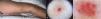
Physical examinationExamination revealed the presence of an 8-mm erythematous, elastic papule in diameter. Dermoscopy showed a homogeneous erythematoviolaceous background with multiple corkscrew vessels of various calibers scattered throughout the lesion, intermingled with several irregular linear vessels (Fig. 1B and C).
Ancillary tests
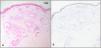
Given this vascular dermoscopic pattern and the differential diagnoses of amelanotic melanoma/melanoma metastasis vs a vascular lesion, an excisional biopsy was performed. Histology revealed a well-circumscribed, nonencapsulated dermal proliferation of thick-walled vessels (Fig. 2A). Melan-A immunohistochemistry was negative within the tumor (Fig. 2B).

What is your diagnosis?
DiagnosisArteriovenous tumor.
Treatment and follow-upThe patient has remained clinically stable, with no signs of local recurrence 1 year after excision.
DiscussionArteriovenous tumor – also called arteriovenous hemangioma or acral arteriovenous tumor – is a benign acquired neoplasm of unknown origin.1 It typically begins in middle age as a single, asymptomatic papule 0.5–1cm in size, with a predilection for the skin of the face or extremities.1,2 Histologically, it consists of well-demarcated, nonencapsulated proliferations of thick-walled dermal vessels.1 It is considered underdiagnosed because its clinical presentation is nonspecific.1,2 Dermoscopy can be crucial for including this entity in the differential diagnosis of compatible lesions; the most frequent dermoscopic findings are non-arborizing telangiectasias, whitish structures on a homogeneously erythematous or violaceous background, and frequent absence of vascular lagoons.1,2 However, up to the present case, there were no reports in the literature of an arteriovenous tumor showing corkscrew vessels.
Corkscrew vessels are helical/spiral vascular dermoscopic structures that, in more than 80% of cases, are associated with melanoma metastases or nodular/desmoplastic melanomas.3 They have also been reported in other tumors relevant to this case's differential diagnosis, such as basal cell carcinoma4 and acquired elastotic hemangioma.3 The latter, first described by Requena et al. in 2002, is a benign, slow-growing vascular tumor that can resemble arteriovenous tumor, typically presenting as small solitary erythematoviolaceous papules/plaques on photoexposed areas of elderly or middle-aged women.3,5 Histologically – unlike arteriovenous tumor – it features a band-like proliferation of capillaries in the papillary dermis arranged horizontally parallel to the epidermis, along with marked dermal elastosis.3,5,6
Because it is entirely benign, arteriovenous tumor does not require treatment or follow-up. Nevertheless, since it often raises diagnostic doubts with other entities – including malignant ones – it is commonly excised for diagnostic purposes.3
In conclusion, we report the first case in the literature of an arteriovenous tumor with the dermoscopic finding of corkscrew vessels. This case underscores the importance of considering alternatives to melanoma when confronted with this dermoscopic sign.
Conflicts of interestNone declared.


