


Pyoderma gangrenosum (PG) is a neutrophilic dermatosis (ND) that is frequently associated with systemic diseases. Its association with systemic lupus erythematosus (SLE) is, however, exceptionally rare. We describe a case in which PG preceded the diagnosis of SLE and highlight therapeutic aspects.
A 59-year-old woman with a past medical history of hypertension, type 2 diabetes mellitus, and bipolar disorder presented to the emergency department with a painful lesion on the right leg of 2 weeks’ duration which, despite levofloxacin and amoxicillin–clavulanate, enlarged and ulcerated. The history did not identify other relevant systemic symptoms.
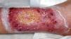
On examination, the patient was dehydrated, hypotensive (80/55mmHg), and tachypneic (31breaths/min), with a single ulcerated plaque showing depressed erythematous–violaceous borders (Fig. 1). Laboratory tests showed hemoglobin 11.4g/dL, leukocytes 11.2×10³/μL, neutrophils 9.6×10³/μL, platelets 414×103/μL, and C-reactive protein 12.6mg/dL. CT revealed marked edema of subcutaneous and muscle tissue. She was admitted and started on IV normal saline and empiric antibiotics with piperacillin–tazobactam plus gentamicin.

The following day she developed chest pain. Repeat labs showed hemoglobin 8.5g/dL, leukocytes 0.3×10³/μL, neutrophils 0.1×10³/μL, platelets 267×103/μL, D-dimer 35,336ng/mL, and a positive direct Coombs test. Peripheral smear, CT angiography, and transthoracic echocardiogram were normal. A biopsy was taken from the lesion edge, and a single 100mg IV bolus of methylprednisolone was administered along with filgrastim.
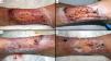
On hospital day 2, prednisone 1mg/kg/day was begun, and daily wound care with wet compresses and a barrier cream containing copper and zinc sulfate was performed (Fig. 2). Notable improvement was observed from day 4 and continued during hospitalization. However, on day 5, thrombocytopenia emerged.

Clinical evolution of the lesion. Daily wound care consisted of wet compresses with zinc, copper, and aluminum–potassium sulfates, followed by careful mechanical debridement of necrotic tissue and application of collagenase to remaining slough. Islands of spared and surrounding skin were protected with a barrier cream containing copper and zinc sulfate, then covered with a silver alginate dressing and bandage. Clinical appearance at 48h (A) and 96h (B) after initiation of the comprehensive approach shows clear improvement in erythema, edema, and ulceration. By day 5 (C) the response remained favorable and progressive. At day 10 (D), re-epithelialization was evident both concentrically from lesion borders and eccentrically from islands of spared skin inward over debrided areas.
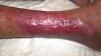
Histopathology showed a dense neutrophilic infiltrate in the dermis. PAS stain was negative. Antinuclear antibodies (ANA) were elevated (titer 1:1280) with anti-double-stranded DNA antibodies 83IU/mL. All microbiologic cultures obtained during hospitalization tested negative, and antibiotics were discontinued on day 10. On day 15, due to progressive improvement, she was discharged on hydroxychloroquine and a tapering course of prednisone. Despite complete re-epithelialization of the lesion at the 1-month follow-up (Fig. 3), she required readmission for a flare of lupus nephritis, which confirmed the SLE diagnosis. Therapy was adjusted and belimumab initiated, with no PG recurrences at 1-year follow-up.

We presented an unusual case in which PG preceded fulfillment of EULAR/ACR criteria for SLE diagnosis.1 In a review by Magdoud et al. of 25 PG–SLE cases, 72% received the SLE diagnosis before PG onset, whereas in 12% PG preceded SLE diagnosis.2 Additionally, patients with PG and SLE tend to be younger than those with PG without SLE. Nevertheless, this association appears not to significantly impact SLE prognosis and seems independent of disease activity.2
Successful management of PG associated with SLE involves reducing inflammation and optimizing wound healing while simultaneously treating the underlying disease. Our literature review (Table 1) incorporates therapeutic aspects not previously addressed in depth. First, we found heterogeneity in both treatments employed and clinical responses.2–8 The most widely used drugs were prednisone (72.7%) and cyclosporine (42.4%). Although complete remission was achieved in most cases (75.7%), relapses occurred up to 2 years later, underscoring the need for continued clinical surveillance. Second, data on topical treatment are limited. Angiogenesis – fundamental for wound healing – is largely regulated by vascular endothelial growth factor (VEGF). It has been suggested that copper may positively influence VEGF expression via pathways similar to tissue hypoxia, and that topical copper sulfate might accelerate contraction and closure of dermal wounds.9 Conversely, although aggressive surgical debridement is generally discouraged in PG, in our case we used a strategy of meticulous, sequential selective debridement of necrotic tissue. Importantly, the scarcity of detailed information hampers meaningful comparisons among therapeutic approaches and the formulation of precise management recommendations to treat these injuries.
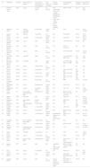
Cases of PG-related SLE.
| No. | Reference | Sex/age | Site (number of lesions) | Timing of PG onset relative to SLE diagnosis | SLE activity at PG onset | Topical treatment | Pharmacologic treatment | Response (months) | Recurrence (follow-up) |
|---|---|---|---|---|---|---|---|---|---|
| 1 | Present clinical case | F/59 | Leg (1) | 1 month before | No activity | Wet compresses with Cu/Zn sulfates, debridement, collagenase, barrier cream with Cu/Zn sulfates, silver alginate dressing | MPD-IV; PDN | CR (1) | No (1 year) |
| 2 | Magdoud [2019] | F/43 | Thighs, shoulders, hands (6) | 4 years after | Active signs | ND | PDN | CR (1) | No (6 months) |
| 3 | Lebrun [2018] | F/32 | Face (3) | 1 year after | No activity | ND | TCS; MINO | CR (6) | No (3 years) |
| 4 | Lebrun [2018] | F/37 | Leg (MP) | 10 years after | Active signs | ND | PDN; MTX | CR (1) | No (3 years) |
| 5 | Gonzalez-Moreno [2015] | M/46 | Leg (1) | After | Active signs | ND | TCS; PDN; CsA | CR (3) | ND |
| 6 | Gonzalez-Moreno [2015] | F/63 | Leg (1) | After | No activity | ND | TCS; IV tacrolimus | ND | ND |
| 7 | Gonzalez-Moreno [2015] | F/42 | Leg (1) | After | No activity | ND | TCS; PDN; AZA | CR (3) | Yes (2 years) |
| 8 | Gonzalez-Moreno [2015] | F/46 | Leg (MP) | After | No activity | ND | TCS; CsA; AZA; MMF; IVIG | CR (ND) | Yes (ND) |
| 9 | Gonzalez-Moreno [2015] | M/36 | Foot (MP) | After | Active signs | ND | ILC; PDN; AZA; DDS; CF; MMF | CR (ND) | ND |
| 10 | Hamzi [2013] | F/35 | Leg (1) | 18 years after | No activity | ND | ND | ND | ND |
| 11 | Canas [2010] | ND/48 | Leg (1) | 2 years after | ND | ND | PDN; CsA; ASA; WF | CR (ND) | ND |
| 12 | Canas [2010] | ND/28 | Leg (1) | 4 years after | Active signs | ND | PDN; CsA; ASA; WF | CR (ND) | ND |
| 13 | Canas [2010] | ND/50 | Foot (1) | Simultaneous | ND | ND | PDN; CsA; ASA; WF | CR (ND) | ND |
| 14 | Husein-ElAhmed [2010] | M/36 | Foot (1) | 8 years after | No activity | ND | MMF | CR (1) | No (ND) |
| 15 | Masatlioglu [2009] | F/35 | Leg (1) | Simultaneous | Active signs | ND | PDN; HCQ | CR (1) | ND |
| 16 | Masatlioglu [2009] | F/47 | Thigh (1) | 10 months before | Active signs | ND | CsA; TCS | CR (2) | No (6 months) |
| 17 | Hind [2008] | F/1 | Face, upper and lower limbs (MP) | Simultaneous | Active signs | ND | MPD-IV; PDN; AZT; CsA; IVIG; TLD; IFX | NR | Died |
| 18 | Reddy [2007] | M/34 | Legs (MP) | After | Active signs | ND | PDN; AZT | CR (1) | No (3 months) |
| 19 | Waldman [2005] | F/35 | Lower limbs (MP) | 5 years before | ND | ND | PDN; CsA; MMF | CR (11) | Yes (8 months) |
| 20 | Sakamoto [2002] | F/55 | Trunk/shoulders (MP) | 3 years after | Active signs | ND | CsA | CR (ND) | No (ND) |
| 21 | Schmid [1998] | F/64 | Legs (2) | 11 years after | Active signs | ND | PDN; CsA | NR | Died |
| 22 | Holbrook [1996] | F/57 | Leg (1) | 2 years after | No activity | ND | MPD-IV; PDN; MTX | CR (6) | No (ND) |
| 23 | Roger [1993] | F/25 | Foot (1) | 1 month after | Active signs | Dextranomer followed by sterile hydrocolloid | MPD-IV; CF; PDN | CR (4) | No (10 months) |
| 24 | Hostetler [1993] | F/27 | Trunk, buttocks, knees (MP) | 13 years after | No activity | ND | ND | ND | ND |
| 25 | Pinto [1991] | F/35 | Leg (1) | 15 years after | Active signs | ND | MPD-IV; PDN | CR (1) | No (4 months) |
| 26 | Peterson [1984] | F/48 | Intergluteal and inguinal folds (6) | Simultaneous | Active signs | ND | TCS | CR (2) | No (ND) |
| 27 | Olson [1971] | F/15 | Leg (MP) | 1 year before | ND | ND | PDN | ND | Yes (2 years) |
| 28 | Hania [2014] | F/53 | Leg (2) | 17 years after | No activity | ND | PDN | CR (6) | ND |
| 29 | Teoh [2021] | M/35 | Scrotum, legs (MP) | Simultaneous | Active signs | ND | PDN; HCQ; MTX; cyanocobalamin | CR (1) | ND |
| 30 | Beynon [2017] | F/57 | Leg (1) | After | ND | Hydrofiber and hydrocolloid dressings | MPD-IV; PDN; TLD; CsA; MMF; ANAK | PR (11) | ND |
| 31 | Choi [2018] | F/61 | Leg (1) | 8 years after | No activity | ND | PDN; CsA | CR (6) | No (1 year) |
| 32 | Ibrahim [2021] | F/55 | Leg (1) | After | Active signs | ND | PDN; DDS; MTX; MMF; CsA; IVIG | PR (24) | ND |
| 33 | Ahmadi [2023] | F/40 | Leg (1) | After | No activity | Saline solution, mupirocin, petrolatum gauze | PDN; MPD-IV; CsA; AZT; intralesional IFX | CR (3) | ND |
ASA: acetylsalicylic acid; ANAK: anakinra; AZA: azathioprine; CF: cyclophosphamide; CsA: cyclosporine; Cu/Zn: copper/zinc; DDS: dapsone; HCQ: hydroxychloroquine; ILC: intralesional corticosteroids; IFX: infliximab; IVIG: IV immunoglobulins; SLE: systemic lupus erythematosus; M: male; F: female; MINO: minocycline; MMF: mycophenolate mofetil; MP: multiple; MPD-IV: IV methylprednisolone; MTX: methotrexate; ND: not available; PDN: prednisone; PG: pyoderma gangrenosum; CR: complete response; PR: partial response; TCS: topical corticosteroids; TLD: thalidomide; WF: warfarin.
In conclusion, the heterogeneity of therapeutic approaches to SLE-related PG emphasizes the need for individualized treatment protocols, highlighting the importance of a deeper understanding of topical and systemic therapies to optimize management.
Conflicts of interestNone declared.


