array:24 [
  "pii" => "S157821901400256X"
  "issn" => "15782190"
  "doi" => "10.1016/j.adengl.2014.09.015"
  "estado" => "S300"
  "fechaPublicacion" => "2014-11-01"
  "aid" => "984"
  "copyright" => "Elsevier España, S.L.U. and AEDV"
  "copyrightAnyo" => "2013"
  "documento" => "simple-article"
  "crossmark" => 0
  "subdocumento" => "cor"
  "cita" => "Actas Dermosifiliogr. 2014;105:878-9"
  "abierto" => array:3 [
    "ES" => true
    "ES2" => true
    "LATM" => true
  ]
  "gratuito" => true
  "lecturas" => array:2 [
    "total" => 1469
    "formatos" => array:3 [
      "EPUB" => 39
      "HTML" => 992
      "PDF" => 438
    ]
  ]
  "Traduccion" => array:1 [
    "es" => array:19 [
      "pii" => "S0001731014001033"
      "issn" => "00017310"
      "doi" => "10.1016/j.ad.2014.03.001"
      "estado" => "S300"
      "fechaPublicacion" => "2014-11-01"
      "aid" => "984"
      "copyright" => "Elsevier España, S.L. y AEDV"
      "documento" => "simple-article"
      "crossmark" => 0
      "subdocumento" => "cor"
      "cita" => "Actas Dermosifiliogr. 2014;105:878-9"
      "abierto" => array:3 [
        "ES" => true
        "ES2" => true
        "LATM" => true
      ]
      "gratuito" => true
      "lecturas" => array:2 [
        "total" => 3128
        "formatos" => array:3 [
          "EPUB" => 1
          "HTML" => 1948
          "PDF" => 1179
        ]
      ]
      "es" => array:11 [
        "idiomaDefecto" => true
        "cabecera" => "<span class="elsevierStyleTextfn">Carta cient&#237;fico-cl&#237;nica</span>"
        "titulo" => "Tumor primitivo polipoide de c&#233;lulas granulares"
        "tienePdf" => "es"
        "tieneTextoCompleto" => "es"
        "paginas" => array:1 [
          0 => array:2 [
            "paginaInicial" => "878"
            "paginaFinal" => "879"
          ]
        ]
        "titulosAlternativos" => array:1 [
          "en" => array:1 [
            "titulo" => "Primitive Polypoid Granular-Cell Tumor"
          ]
        ]
        "contieneTextoCompleto" => array:1 [
          "es" => true
        ]
        "contienePdf" => array:1 [
          "es" => true
        ]
        "resumenGrafico" => array:2 [
          "original" => 0
          "multimedia" => array:7 [
            "identificador" => "fig0005"
            "etiqueta" => "Figura 1"
            "tipo" => "MULTIMEDIAFIGURA"
            "mostrarFloat" => true
            "mostrarDisplay" => false
            "figura" => array:1 [
              0 => array:4 [
                "imagen" => "gr1.jpeg"
                "Alto" => 1811
                "Ancho" => 1583
                "Tamanyo" => 683627
              ]
            ]
            "descripcion" => array:1 [
              "es" => "<p id="spar0005" class="elsevierStyleSimplePara elsevierViewall">H-E &#215;<span class="elsevierStyleHsp" style=""></span>10&#46; Tumoraci&#243;n localizada en la dermis superficial y media&#44; rodeada de un collarete epitelial&#46;</p>"
            ]
          ]
        ]
        "autores" => array:1 [
          0 => array:2 [
            "autoresLista" => "M&#46;T&#46; L&#243;pez-Villaescusa, M&#46; Rodr&#237;guez-V&#225;zquez, M&#46; Garc&#237;a-Arpa, R&#46; Garc&#237;a-Angel"
            "autores" => array:4 [
              0 => array:2 [
                "nombre" => "M&#46;T&#46;"
                "apellidos" => "L&#243;pez-Villaescusa"
              ]
              1 => array:2 [
                "nombre" => "M&#46;"
                "apellidos" => "Rodr&#237;guez-V&#225;zquez"
              ]
              2 => array:2 [
                "nombre" => "M&#46;"
                "apellidos" => "Garc&#237;a-Arpa"
              ]
              3 => array:2 [
                "nombre" => "R&#46;"
                "apellidos" => "Garc&#237;a-Angel"
              ]
            ]
          ]
        ]
      ]
      "idiomaDefecto" => "es"
      "Traduccion" => array:1 [
        "en" => array:9 [
          "pii" => "S157821901400256X"
          "doi" => "10.1016/j.adengl.2014.09.015"
          "estado" => "S300"
          "subdocumento" => ""
          "abierto" => array:3 [
            "ES" => true
            "ES2" => true
            "LATM" => true
          ]
          "gratuito" => true
          "lecturas" => array:1 [
            "total" => 0
          ]
          "idiomaDefecto" => "en"
          "EPUB" => "https://multimedia.elsevier.es/PublicationsMultimediaV1/item/epub/S157821901400256X?idApp=UINPBA000044"
        ]
      ]
      "EPUB" => "https://multimedia.elsevier.es/PublicationsMultimediaV1/item/epub/S0001731014001033?idApp=UINPBA000044"
      "url" => "/00017310/0000010500000009/v1_201410280121/S0001731014001033/v1_201410280121/es/main.assets"
    ]
  ]
  "itemSiguiente" => array:19 [
    "pii" => "S1578219014002571"
    "issn" => "15782190"
    "doi" => "10.1016/j.adengl.2014.03.003"
    "estado" => "S300"
    "fechaPublicacion" => "2014-11-01"
    "aid" => "991"
    "copyright" => "Elsevier Espa&#241;a&#44; S&#46;L&#46;U&#46; and AEDV"
    "documento" => "simple-article"
    "crossmark" => 0
    "subdocumento" => "cor"
    "cita" => "Actas Dermosifiliogr. 2014;105:879-82"
    "abierto" => array:3 [
      "ES" => true
      "ES2" => true
      "LATM" => true
    ]
    "gratuito" => true
    "lecturas" => array:2 [
      "total" => 1242
      "formatos" => array:3 [
        "EPUB" => 40
        "HTML" => 815
        "PDF" => 387
      ]
    ]
    "en" => array:11 [
      "idiomaDefecto" => true
      "cabecera" => "<span class="elsevierStyleTextfn">Case and Research Letters</span>"
      "titulo" => "Dermal Leiomyosarcoma at the End of the Left Eyebrow"
      "tienePdf" => "en"
      "tieneTextoCompleto" => "en"
      "paginas" => array:1 [
        0 => array:2 [
          "paginaInicial" => "879"
          "paginaFinal" => "882"
        ]
      ]
      "titulosAlternativos" => array:1 [
        "es" => array:1 [
          "titulo" => "Leiomiosarcoma d&#233;rmico en la cola de la ceja izquierda"
        ]
      ]
      "contieneTextoCompleto" => array:1 [
        "en" => true
      ]
      "contienePdf" => array:1 [
        "en" => true
      ]
      "resumenGrafico" => array:2 [
        "original" => 0
        "multimedia" => array:7 [
          "identificador" => "fig0010"
          "etiqueta" => "Figure 2"
          "tipo" => "MULTIMEDIAFIGURA"
          "mostrarFloat" => true
          "mostrarDisplay" => false
          "figura" => array:1 [
            0 => array:4 [
              "imagen" => "gr2.jpeg"
              "Alto" => 1141
              "Ancho" => 1401
              "Tamanyo" => 328602
            ]
          ]
          "descripcion" => array:1 [
            "en" => "<p id="spar0010" class="elsevierStyleSimplePara elsevierViewall">A&#44; Panoramic microscopic image shows a neoplasm with an expansive growth pattern&#44; which does not affect the resection margin &#40;hematoxylin-eosin&#44; original magnification &#215;4&#41;&#46; B&#44; The neoplasm is composed of spindle cells arranged in fascicles with marked atypia and abundant mitotic figures &#40;hematoxylin-eosin&#44; original magnification &#215;40&#41;&#46; C&#44; Expression of smooth muscle actin &#40;Papanicolaou&#44; original magnification &#215;40&#41;&#46; D&#44; The proliferative index &#40;Papanicolaou&#44; Ki-67&#41; is 20&#37;&#46;</p>"
          ]
        ]
      ]
      "autores" => array:1 [
        0 => array:2 [
          "autoresLista" => "A&#46;D&#46; Lozano Salazar, A&#46; M&#225;rquez Garc&#237;a, I&#46; Ortega Medina, J&#46;J&#46; R&#237;os-Mart&#237;n"
          "autores" => array:4 [
            0 => array:2 [
              "nombre" => "A&#46;D&#46;"
              "apellidos" => "Lozano Salazar"
            ]
            1 => array:2 [
              "nombre" => "A&#46;"
              "apellidos" => "M&#225;rquez Garc&#237;a"
            ]
            2 => array:2 [
              "nombre" => "I&#46;"
              "apellidos" => "Ortega Medina"
            ]
            3 => array:2 [
              "nombre" => "J&#46;J&#46;"
              "apellidos" => "R&#237;os-Mart&#237;n"
            ]
          ]
        ]
      ]
    ]
    "idiomaDefecto" => "en"
    "Traduccion" => array:1 [
      "es" => array:9 [
        "pii" => "S0001731014001823"
        "doi" => "10.1016/j.ad.2014.03.003"
        "estado" => "S300"
        "subdocumento" => ""
        "abierto" => array:3 [
          "ES" => true
          "ES2" => true
          "LATM" => true
        ]
        "gratuito" => true
        "lecturas" => array:1 [
          "total" => 0
        ]
        "idiomaDefecto" => "es"
        "EPUB" => "https://multimedia.elsevier.es/PublicationsMultimediaV1/item/epub/S0001731014001823?idApp=UINPBA000044"
      ]
    ]
    "EPUB" => "https://multimedia.elsevier.es/PublicationsMultimediaV1/item/epub/S1578219014002571?idApp=UINPBA000044"
    "url" => "/15782190/0000010500000009/v1_201410280115/S1578219014002571/v1_201410280115/en/main.assets"
  ]
  "itemAnterior" => array:19 [
    "pii" => "S1578219014002558"
    "issn" => "15782190"
    "doi" => "10.1016/j.adengl.2014.09.014"
    "estado" => "S300"
    "fechaPublicacion" => "2014-11-01"
    "aid" => "969"
    "copyright" => "Elsevier Espa&#241;a&#44; S&#46;L&#46;U&#46; and AEDV"
    "documento" => "simple-article"
    "crossmark" => 0
    "subdocumento" => "cor"
    "cita" => "Actas Dermosifiliogr. 2014;105:876-8"
    "abierto" => array:3 [
      "ES" => true
      "ES2" => true
      "LATM" => true
    ]
    "gratuito" => true
    "lecturas" => array:2 [
      "total" => 1185
      "formatos" => array:3 [
        "EPUB" => 39
        "HTML" => 891
        "PDF" => 255
      ]
    ]
    "en" => array:11 [
      "idiomaDefecto" => true
      "cabecera" => "<span class="elsevierStyleTextfn">Case and Research Letters</span>"
      "titulo" => "Dermatomyositis-Like Eruption Associated With Hydroxyurea Therapy&#58; A Premalignant Condition&#63;"
      "tienePdf" => "en"
      "tieneTextoCompleto" => "en"
      "paginas" => array:1 [
        0 => array:2 [
          "paginaInicial" => "876"
          "paginaFinal" => "878"
        ]
      ]
      "titulosAlternativos" => array:1 [
        "es" => array:1 [
          "titulo" => "Erupci&#243;n dermatomiositis-<span class="elsevierStyleItalic">like</span> asociada al tratamiento con hidroxiurea&#58; &#191;una entidad premaligna&#63;"
        ]
      ]
      "contieneTextoCompleto" => array:1 [
        "en" => true
      ]
      "contienePdf" => array:1 [
        "en" => true
      ]
      "resumenGrafico" => array:2 [
        "original" => 0
        "multimedia" => array:7 [
          "identificador" => "fig0005"
          "etiqueta" => "Figure 1"
          "tipo" => "MULTIMEDIAFIGURA"
          "mostrarFloat" => true
          "mostrarDisplay" => false
          "figura" => array:1 [
            0 => array:4 [
              "imagen" => "gr1.jpeg"
              "Alto" => 866
              "Ancho" => 1300
              "Tamanyo" => 225108
            ]
          ]
          "descripcion" => array:1 [
            "en" => "<p id="spar0005" class="elsevierStyleSimplePara elsevierViewall">A&#44; Desquamating erythematous lesions in the periorbital region&#46; B&#44; Erythematous papular lesions in the retroauricular region&#46;</p>"
          ]
        ]
      ]
      "autores" => array:1 [
        0 => array:2 [
          "autoresLista" => "B&#46; de Unamuno-Bustos, R&#46; Ballester-S&#225;nchez, V&#46; Sabater Marco, J&#46;J&#46; Vilata-Corell"
          "autores" => array:4 [
            0 => array:2 [
              "nombre" => "B&#46;"
              "apellidos" => "de Unamuno-Bustos"
            ]
            1 => array:2 [
              "nombre" => "R&#46;"
              "apellidos" => "Ballester-S&#225;nchez"
            ]
            2 => array:2 [
              "nombre" => "V&#46;"
              "apellidos" => "Sabater Marco"
            ]
            3 => array:2 [
              "nombre" => "J&#46;J&#46;"
              "apellidos" => "Vilata-Corell"
            ]
          ]
        ]
      ]
    ]
    "idiomaDefecto" => "en"
    "Traduccion" => array:1 [
      "es" => array:9 [
        "pii" => "S0001731014000593"
        "doi" => "10.1016/j.ad.2014.02.003"
        "estado" => "S300"
        "subdocumento" => ""
        "abierto" => array:3 [
          "ES" => true
          "ES2" => true
          "LATM" => true
        ]
        "gratuito" => true
        "lecturas" => array:1 [
          "total" => 0
        ]
        "idiomaDefecto" => "es"
        "EPUB" => "https://multimedia.elsevier.es/PublicationsMultimediaV1/item/epub/S0001731014000593?idApp=UINPBA000044"
      ]
    ]
    "EPUB" => "https://multimedia.elsevier.es/PublicationsMultimediaV1/item/epub/S1578219014002558?idApp=UINPBA000044"
    "url" => "/15782190/0000010500000009/v1_201410280115/S1578219014002558/v1_201410280115/en/main.assets"
  ]
  "en" => array:14 [
    "idiomaDefecto" => true
    "cabecera" => "<span class="elsevierStyleTextfn">Case and Research Letters</span>"
    "titulo" => "Primitive Polypoid Granular-Cell Tumor"
    "tieneTextoCompleto" => true
    "paginas" => array:1 [
      0 => array:2 [
        "paginaInicial" => "878"
        "paginaFinal" => "879"
      ]
    ]
    "autores" => array:1 [
      0 => array:4 [
        "autoresLista" => "M&#46;T&#46; L&#243;pez-Villaescusa, M&#46; Rodr&#237;guez-V&#225;zquez, M&#46; Garc&#237;a-Arpa, R&#46; Garc&#237;a-Angel"
        "autores" => array:4 [
          0 => array:4 [
            "nombre" => "M&#46;T&#46;"
            "apellidos" => "L&#243;pez-Villaescusa"
            "email" => array:1 [
              0 => "lopezvillaescusa&#64;hotmail&#46;com"
            ]
            "referencia" => array:2 [
              0 => array:2 [
                "etiqueta" => "<span class="elsevierStyleSup">a</span>"
                "identificador" => "aff0005"
              ]
              1 => array:2 [
                "etiqueta" => "<span class="elsevierStyleSup">&#42;</span>"
                "identificador" => "cor0005"
              ]
            ]
          ]
          1 => array:3 [
            "nombre" => "M&#46;"
            "apellidos" => "Rodr&#237;guez-V&#225;zquez"
            "referencia" => array:1 [
              0 => array:2 [
                "etiqueta" => "<span class="elsevierStyleSup">a</span>"
                "identificador" => "aff0005"
              ]
            ]
          ]
          2 => array:3 [
            "nombre" => "M&#46;"
            "apellidos" => "Garc&#237;a-Arpa"
            "referencia" => array:1 [
              0 => array:2 [
                "etiqueta" => "<span class="elsevierStyleSup">b</span>"
                "identificador" => "aff0010"
              ]
            ]
          ]
          3 => array:3 [
            "nombre" => "R&#46;"
            "apellidos" => "Garc&#237;a-Angel"
            "referencia" => array:1 [
              0 => array:2 [
                "etiqueta" => "<span class="elsevierStyleSup">c</span>"
                "identificador" => "aff0015"
              ]
            ]
          ]
        ]
        "afiliaciones" => array:3 [
          0 => array:3 [
            "entidad" => "Servicio de Dermatolog&#237;a&#44; Complejo Hospitalario Universitario de Albacete&#44; Albacete&#44; Spain"
            "etiqueta" => "a"
            "identificador" => "aff0005"
          ]
          1 => array:3 [
            "entidad" => "Servicio de Dermatolog&#237;a&#44; Hospital de Ciudad Real&#44; Ciudad Real&#44; Spain"
            "etiqueta" => "b"
            "identificador" => "aff0010"
          ]
          2 => array:3 [
            "entidad" => "Servicio de Anatom&#237;a Patol&#243;gica&#44; Complejo Hospitalario Universitario de Albacete&#44; Albacete&#44; Spain"
            "etiqueta" => "c"
            "identificador" => "aff0015"
          ]
        ]
        "correspondencia" => array:1 [
          0 => array:3 [
            "identificador" => "cor0005"
            "etiqueta" => "&#8270;"
            "correspondencia" => "Corresponding author&#46;"
          ]
        ]
      ]
    ]
    "titulosAlternativos" => array:1 [
      "es" => array:1 [
        "titulo" => "Tumor primitivo polipoide de c&#233;lulas granulares"
      ]
    ]
    "resumenGrafico" => array:2 [
      "original" => 0
      "multimedia" => array:7 [
        "identificador" => "fig0010"
        "etiqueta" => "Figure 2"
        "tipo" => "MULTIMEDIAFIGURA"
        "mostrarFloat" => true
        "mostrarDisplay" => false
        "figura" => array:1 [
          0 => array:4 [
            "imagen" => "gr2.jpeg"
            "Alto" => 586
            "Ancho" => 950
            "Tamanyo" => 221167
          ]
        ]
        "descripcion" => array:1 [
          "en" => "<p id="spar0010" class="elsevierStyleSimplePara elsevierViewall">Cells with a polygonal morphology&#44; abundant granular eosinophilic cytoplasm&#44; and large vesicular nuclei&#46; The cells are arranged in an interlinked fascicular pattern&#46; Hematoxylin-eosin&#44; original magnification &#215;40&#46;</p>"
        ]
      ]
    ]
    "textoCompleto" => "<span class="elsevierStyleSections"><p id="par0005" class="elsevierStylePara elsevierViewall">In contrast to conventional granular cell tumor &#40;Abrikossoff tumor&#41;&#44; primitive polypoid granular cell tumor was first identified by LeBoit et al&#46;<a class="elsevierStyleCrossRef" href="#bib0005"><span class="elsevierStyleSup">1</span></a> in 1991 and subsequently endorsed by Chaudhry and Calonje<a class="elsevierStyleCrossRef" href="#bib0010"><span class="elsevierStyleSup">2</span></a> as a dermal tumor of granular cells of non-neural origin&#46; The tumor has a polypoid morphology and presents numerous mitoses&#44; cytologic atypia&#44; and a primitive immunophenotype&#46; We present a new case and review the characteristics of this rare and poorly known tumor&#46;</p><p id="par0010" class="elsevierStylePara elsevierViewall">Our patient was a woman aged 44 years&#44; with no past medical or family history of interest&#46; She consulted for an asymptomatic lesion that had appeared 4 months earlier at the right nasolabial angle&#46; Physical examination revealed a hard&#44; polypoid lesion of 0&#46;3<span class="elsevierStyleHsp" style=""></span>mm diameter&#44; with a translucent surface&#46; With a possible diagnosis of fibrous papule&#44; milium cyst&#44; or adnexal tumor &#40;trichodiscoma&#41;&#44; the lesion was excised&#46; Histopathology revealed a circumscribed proliferation of cells in the superficial and mid dermis&#44; surrounded by an epithelial collarette &#40;<a class="elsevierStyleCrossRef" href="#fig0005">Fig&#46; 1</a>&#41;&#46; The cells&#44; arranged in an interlinked fascicular pattern&#44; had a poligonal morphology with abundant&#44; granular eosinophilic cytoplasm and large vesicular nuclei &#40;<a class="elsevierStyleCrossRef" href="#fig0010">Fig&#46; 2</a>&#41;&#46; Mitotic figures were present&#46; No ulceration or necrosis was observed&#46; Immunohistochemistry was positive for CD68 and negative for AE1-AE3&#44; S-100&#44; Melan A&#44; CD34&#44; desmin&#44; actin&#44; and smooth muscle&#46; On the basis of these findings we made a diagnosis of primitive polypoid granular cell tumor&#46;</p><elsevierMultimedia ident="fig0005"></elsevierMultimedia><elsevierMultimedia ident="fig0010"></elsevierMultimedia><p id="par0015" class="elsevierStylePara elsevierViewall">Granular cells can be found in a varied group of tumors and reflect the intracytoplasmic accumulation of lysosomes and other components of the Golgi aparatus&#46; Traditional and conventional nomenclature makes reference to the cutaneous and mucosal granular cell tumor&#44; known as Abrikossoff tumor&#44; a benign neoplasm of neural origin derived from Schwann cells&#46;<a class="elsevierStyleCrossRef" href="#bib0015"><span class="elsevierStyleSup">3</span></a> However&#44; other granular cell tumors of non-neural origin exist&#44; including the congenital gingival granular cell tumor and the primitive polypoid granular cell tumor&#46; In addition&#44; numerous tumors can present granular cells&#44; including myogenic tumors&#44; melanocytic lesions&#44; dermatofibroma&#44; dermatofibrosarcoma protuberans&#44; basal cell carcinoma&#44; atypical fibroxanthoma&#44; angiosarcoma&#44; fibrous papule&#44; ameloblastoma&#44; and adnexal tumors with eccrine or apocrine differentiation&#46;<a class="elsevierStyleCrossRef" href="#bib0020"><span class="elsevierStyleSup">4</span></a></p><p id="par0020" class="elsevierStylePara elsevierViewall">Primitive polypoid granular cell tumor is a rare neoplasm or uncertain origin that affects middle-aged adults&#46; It usually arises on the trunk or limbs as a polypoid or elevated lesion with a smooth surface&#59; its size can be variable&#46; The distinctive characteristic that differentiates it from conventional granular cell tumor is its histopathology&#44; which shows a tumor in the mid dermis that is clearly surrounded by an epidermal collarette&#59; the tumor is formed of large polygonal&#44; round or spindle-shaped cells with marked nuclear pleomorphism&#44; an elongated nucleus&#44; abundant eosinophilic cytoplasm containing fine granules&#44; and mitotic activity of around 1 to 3 mitoses per mm&#44;<a class="elsevierStyleCrossRef" href="#bib0010"><span class="elsevierStyleSup">2</span></a> usually with no atypia and minimal or absent epidermal hyperplasia&#46; These histopathology findings satisfy the criteria proposed by Enzinger and Weiss for the classification of malignant or atypical granular cell tumor&#44; but the tumors can be differentiated by immunohistochemistry&#44; as primitive polypoid granular cell tumor is usually negative for S-100 and positive for CD68 and neuron-specific enolase&#46;<a class="elsevierStyleCrossRef" href="#bib0025"><span class="elsevierStyleSup">5</span></a></p><p id="par0025" class="elsevierStylePara elsevierViewall">Despite the histological characteristics&#44; this is a tumor of low&#8211;grade malignancy&#46; In the series reported there is only 1 case of metastasis&#44; which arose in the cheek 25 months after excision of the lesion and did not present an epidermal collarette on histology&#46;<a class="elsevierStyleCrossRef" href="#bib0030"><span class="elsevierStyleSup">6</span></a></p><p id="par0030" class="elsevierStylePara elsevierViewall">We have presented a new case of primitive polypoid granular cell tumor&#44; a variant that is not clearly distinguished from the conventional tumor&#46; Its atypical histological characteristics allow it to be classified as a new entity and distinguished from granular cell tumor of neural origin&#46;</p></span>"
    "pdfFichero" => "main.pdf"
    "tienePdf" => true
    "NotaPie" => array:1 [
      0 => array:2 [
        "etiqueta" => "&#9734;"
        "nota" => "<p class="elsevierStyleNotepara" id="npar0005">Please cite this article as&#58; L&#243;pez-Villaescusa M&#44; Rodr&#237;guez-V&#225;zquez M&#44; Garc&#237;a-Arpa M&#44; Garc&#237;a-Angel R&#46; Tumor primitivo polipoide de c&#233;lulas granulares&#46; Actas Dermosifiliogr&#46; 2014&#59;105&#58;878&#8211;879&#46;</p>"
      ]
    ]
    "multimedia" => array:2 [
      0 => array:7 [
        "identificador" => "fig0005"
        "etiqueta" => "Figure 1"
        "tipo" => "MULTIMEDIAFIGURA"
        "mostrarFloat" => true
        "mostrarDisplay" => false
        "figura" => array:1 [
          0 => array:4 [
            "imagen" => "gr1.jpeg"
            "Alto" => 1087
            "Ancho" => 950
            "Tamanyo" => 317986
          ]
        ]
        "descripcion" => array:1 [
          "en" => "<p id="spar0005" class="elsevierStyleSimplePara elsevierViewall">Tumor in the superficial and mid dermis&#44; surrounded by an epithelial collarette&#46; Hematoxylin-eosin&#44; original magnification &#215;10&#46;</p>"
        ]
      ]
      1 => array:7 [
        "identificador" => "fig0010"
        "etiqueta" => "Figure 2"
        "tipo" => "MULTIMEDIAFIGURA"
        "mostrarFloat" => true
        "mostrarDisplay" => false
        "figura" => array:1 [
          0 => array:4 [
            "imagen" => "gr2.jpeg"
            "Alto" => 586
            "Ancho" => 950
            "Tamanyo" => 221167
          ]
        ]
        "descripcion" => array:1 [
          "en" => "<p id="spar0010" class="elsevierStyleSimplePara elsevierViewall">Cells with a polygonal morphology&#44; abundant granular eosinophilic cytoplasm&#44; and large vesicular nuclei&#46; The cells are arranged in an interlinked fascicular pattern&#46; Hematoxylin-eosin&#44; original magnification &#215;40&#46;</p>"
        ]
      ]
    ]
    "bibliografia" => array:2 [
      "titulo" => "References"
      "seccion" => array:1 [
        0 => array:2 [
          "identificador" => "bibs0005"
          "bibliografiaReferencia" => array:6 [
            0 => array:3 [
              "identificador" => "bib0005"
              "etiqueta" => "1"
              "referencia" => array:1 [
                0 => array:2 [
                  "contribucion" => array:1 [
                    0 => array:2 [
                      "titulo" => "Primitive polypoid granular-cell tumor and other cutaneous granular-cell neoplasms of apparent nonneural origin"
                      "autores" => array:1 [
                        0 => array:2 [
                          "etal" => false
                          "autores" => array:4 [
                            0 => "E&#46; Philip"
                            1 => "M&#46;D&#46; LeBoit"
                            2 => "J&#46; Ronald"
                            3 => "M&#46;D&#46; Barr"
                          ]
                        ]
                      ]
                    ]
                  ]
                  "host" => array:1 [
                    0 => array:1 [
                      "Revista" => array:6 [
                        "tituloSerie" => "Am J Surg Pathol"
                        "fecha" => "1991"
                        "volumen" => "15"
                        "paginaInicial" => "48"
                        "paginaFinal" => "58"
                        "link" => array:1 [
                          0 => array:2 [
                            "url" => "https://www.ncbi.nlm.nih.gov/pubmed/1985501"
                            "web" => "Medline"
                          ]
                        ]
                      ]
                    ]
                  ]
                ]
              ]
            ]
            1 => array:3 [
              "identificador" => "bib0010"
              "etiqueta" => "2"
              "referencia" => array:1 [
                0 => array:2 [
                  "contribucion" => array:1 [
                    0 => array:2 [
                      "titulo" => "Dermal non-neural granular cell tumor &#40;so-called primitive polypoid granular cell tumor&#41;&#58; A distinctive further delineated in a clinicopathological study of 11 cases"
                      "autores" => array:1 [
                        0 => array:2 [
                          "etal" => false
                          "autores" => array:2 [
                            0 => "I&#46;H&#46; Chaudhry"
                            1 => "E&#46; Caljonje"
                          ]
                        ]
                      ]
                    ]
                  ]
                  "host" => array:1 [
                    0 => array:2 [
                      "doi" => "10.1111/j.1365-2559.2005.02192.x"
                      "Revista" => array:6 [
                        "tituloSerie" => "Histopathology"
                        "fecha" => "2005"
                        "volumen" => "47"
                        "paginaInicial" => "179"
                        "paginaFinal" => "185"
                        "link" => array:1 [
                          0 => array:2 [
                            "url" => "https://www.ncbi.nlm.nih.gov/pubmed/16045779"
                            "web" => "Medline"
                          ]
                        ]
                      ]
                    ]
                  ]
                ]
              ]
            ]
            2 => array:3 [
              "identificador" => "bib0015"
              "etiqueta" => "3"
              "referencia" => array:1 [
                0 => array:2 [
                  "contribucion" => array:1 [
                    0 => array:2 [
                      "titulo" => "Tumor de c&#233;lulas granulares cut&#225;neo&#58; an&#225;lisis cl&#237;nico-patol&#243;gico de treinta y cuatro casos"
                      "autores" => array:1 [
                        0 => array:2 [
                          "etal" => false
                          "autores" => array:5 [
                            0 => "A&#46; Torrijos-Aguilar"
                            1 => "V&#46; Alegre-de Miquel"
                            2 => "G&#46; Pitarch-Bort"
                            3 => "P&#46; Mercader-Garc&#237;a"
                            4 => "J&#46;M&#46; Fortea-Baixauli"
                          ]
                        ]
                      ]
                    ]
                  ]
                  "host" => array:1 [
                    0 => array:1 [
                      "Revista" => array:6 [
                        "tituloSerie" => "Actas Dermosifiliogr"
                        "fecha" => "2009"
                        "volumen" => "100"
                        "paginaInicial" => "126"
                        "paginaFinal" => "132"
                        "link" => array:1 [
                          0 => array:2 [
                            "url" => "https://www.ncbi.nlm.nih.gov/pubmed/19445877"
                            "web" => "Medline"
                          ]
                        ]
                      ]
                    ]
                  ]
                ]
              ]
            ]
            3 => array:3 [
              "identificador" => "bib0020"
              "etiqueta" => "4"
              "referencia" => array:1 [
                0 => array:2 [
                  "contribucion" => array:1 [
                    0 => array:2 [
                      "titulo" => "Cutaneous granular cell tumor&#46; Report of three cases and review of the literature"
                      "autores" => array:1 [
                        0 => array:2 [
                          "etal" => false
                          "autores" => array:2 [
                            0 => "B&#46; Di Martino"
                            1 => "A&#46; Soskin"
                          ]
                        ]
                      ]
                    ]
                  ]
                  "host" => array:1 [
                    0 => array:1 [
                      "Revista" => array:5 [
                        "tituloSerie" => "Our Dermatol Online"
                        "fecha" => "2012"
                        "volumen" => "3"
                        "paginaInicial" => "46"
                        "paginaFinal" => "51"
                      ]
                    ]
                  ]
                ]
              ]
            ]
            4 => array:3 [
              "identificador" => "bib0025"
              "etiqueta" => "5"
              "referencia" => array:1 [
                0 => array:2 [
                  "contribucion" => array:1 [
                    0 => array:2 [
                      "titulo" => "Value of S-100 protein in the diagnosis of soft tissue tumors with particular reference to benign and malignant Schwann cell tumors"
                      "autores" => array:1 [
                        0 => array:2 [
                          "etal" => false
                          "autores" => array:3 [
                            0 => "S&#46;W&#46; Weiss"
                            1 => "J&#46;M&#46; Langloss"
                            2 => "F&#46;M&#46; Enzinger"
                          ]
                        ]
                      ]
                    ]
                  ]
                  "host" => array:1 [
                    0 => array:1 [
                      "Revista" => array:6 [
                        "tituloSerie" => "Lab Invest"
                        "fecha" => "1983"
                        "volumen" => "49"
                        "paginaInicial" => "299"
                        "paginaFinal" => "308"
                        "link" => array:1 [
                          0 => array:2 [
                            "url" => "https://www.ncbi.nlm.nih.gov/pubmed/6310227"
                            "web" => "Medline"
                          ]
                        ]
                      ]
                    ]
                  ]
                ]
              ]
            ]
            5 => array:3 [
              "identificador" => "bib0030"
              "etiqueta" => "6"
              "referencia" => array:1 [
                0 => array:2 [
                  "contribucion" => array:1 [
                    0 => array:2 [
                      "titulo" => "Primitive nonneural granular cell tumors of skin&#58; Clinicopathologic analysis of 13 cases"
                      "autores" => array:1 [
                        0 => array:2 [
                          "etal" => false
                          "autores" => array:2 [
                            0 => "A&#46;J&#46;F&#46; Lazar"
                            1 => "C&#46;D&#46;M&#46; Fletcher"
                          ]
                        ]
                      ]
                    ]
                  ]
                  "host" => array:1 [
                    0 => array:1 [
                      "Revista" => array:6 [
                        "tituloSerie" => "Am J Surg Pathol"
                        "fecha" => "2005"
                        "volumen" => "29"
                        "paginaInicial" => "927"
                        "paginaFinal" => "934"
                        "link" => array:1 [
                          0 => array:2 [
                            "url" => "https://www.ncbi.nlm.nih.gov/pubmed/15958858"
                            "web" => "Medline"
                          ]
                        ]
                      ]
                    ]
                  ]
                ]
              ]
            ]
          ]
        ]
      ]
    ]
  ]
  "idiomaDefecto" => "en"
  "url" => "/15782190/0000010500000009/v1_201410280115/S157821901400256X/v1_201410280115/en/main.assets"
  "Apartado" => array:4 [
    "identificador" => "6157"
    "tipo" => "SECCION"
    "en" => array:2 [
      "titulo" => "Case and Research Letters"
      "idiomaDefecto" => true
    ]
    "idiomaDefecto" => "en"
  ]
  "PDF" => "https://static.elsevier.es/multimedia/15782190/0000010500000009/v1_201410280115/S157821901400256X/v1_201410280115/en/main.pdf?idApp=UINPBA000044&text.app=https://actasdermo.org/"
  "EPUB" => "https://multimedia.elsevier.es/PublicationsMultimediaV1/item/epub/S157821901400256X?idApp=UINPBA000044"
]
Compartir
Información de la revista
Vol. 105. Núm. 9.
Páginas 878-879 (noviembre 2014)
Vol. 105. Núm. 9.
Páginas 878-879 (noviembre 2014)
Case and Research Letters
Acceso a texto completo
Primitive Polypoid Granular-Cell Tumor
Tumor primitivo polipoide de células granulares
Visitas
6161
M.T. López-Villaescusaa,
Autor para correspondencia
lopezvillaescusa@hotmail.com

Corresponding author.
, M. Rodríguez-Vázqueza, M. García-Arpab, R. García-Angelc
a Servicio de Dermatología, Complejo Hospitalario Universitario de Albacete, Albacete, Spain
b Servicio de Dermatología, Hospital de Ciudad Real, Ciudad Real, Spain
c Servicio de Anatomía Patológica, Complejo Hospitalario Universitario de Albacete, Albacete, Spain
Este artículo ha recibido
Información del artículo
Texto completo
Bibliografía
Descargar PDF
Estadísticas
Figuras (2)
Texto completo

In contrast to conventional granular cell tumor (Abrikossoff tumor), primitive polypoid granular cell tumor was first identified by LeBoit et al.1 in 1991 and subsequently endorsed by Chaudhry and Calonje2 as a dermal tumor of granular cells of non-neural origin. The tumor has a polypoid morphology and presents numerous mitoses, cytologic atypia, and a primitive immunophenotype. We present a new case and review the characteristics of this rare and poorly known tumor.

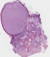
Our patient was a woman aged 44 years, with no past medical or family history of interest. She consulted for an asymptomatic lesion that had appeared 4 months earlier at the right nasolabial angle. Physical examination revealed a hard, polypoid lesion of 0.3mm diameter, with a translucent surface. With a possible diagnosis of fibrous papule, milium cyst, or adnexal tumor (trichodiscoma), the lesion was excised. Histopathology revealed a circumscribed proliferation of cells in the superficial and mid dermis, surrounded by an epithelial collarette (Fig. 1). The cells, arranged in an interlinked fascicular pattern, had a poligonal morphology with abundant, granular eosinophilic cytoplasm and large vesicular nuclei (Fig. 2). Mitotic figures were present. No ulceration or necrosis was observed. Immunohistochemistry was positive for CD68 and negative for AE1-AE3, S-100, Melan A, CD34, desmin, actin, and smooth muscle. On the basis of these findings we made a diagnosis of primitive polypoid granular cell tumor.

Figure 1.

Tumor in the superficial and mid dermis, surrounded by an epithelial collarette. Hematoxylin-eosin, original magnification ×10.

(0.3MB).
Figure 2.

Cells with a polygonal morphology, abundant granular eosinophilic cytoplasm, and large vesicular nuclei. The cells are arranged in an interlinked fascicular pattern. Hematoxylin-eosin, original magnification ×40.

(0.21MB).

Granular cells can be found in a varied group of tumors and reflect the intracytoplasmic accumulation of lysosomes and other components of the Golgi aparatus. Traditional and conventional nomenclature makes reference to the cutaneous and mucosal granular cell tumor, known as Abrikossoff tumor, a benign neoplasm of neural origin derived from Schwann cells.3 However, other granular cell tumors of non-neural origin exist, including the congenital gingival granular cell tumor and the primitive polypoid granular cell tumor. In addition, numerous tumors can present granular cells, including myogenic tumors, melanocytic lesions, dermatofibroma, dermatofibrosarcoma protuberans, basal cell carcinoma, atypical fibroxanthoma, angiosarcoma, fibrous papule, ameloblastoma, and adnexal tumors with eccrine or apocrine differentiation.4

Primitive polypoid granular cell tumor is a rare neoplasm or uncertain origin that affects middle-aged adults. It usually arises on the trunk or limbs as a polypoid or elevated lesion with a smooth surface; its size can be variable. The distinctive characteristic that differentiates it from conventional granular cell tumor is its histopathology, which shows a tumor in the mid dermis that is clearly surrounded by an epidermal collarette; the tumor is formed of large polygonal, round or spindle-shaped cells with marked nuclear pleomorphism, an elongated nucleus, abundant eosinophilic cytoplasm containing fine granules, and mitotic activity of around 1 to 3 mitoses per mm,2 usually with no atypia and minimal or absent epidermal hyperplasia. These histopathology findings satisfy the criteria proposed by Enzinger and Weiss for the classification of malignant or atypical granular cell tumor, but the tumors can be differentiated by immunohistochemistry, as primitive polypoid granular cell tumor is usually negative for S-100 and positive for CD68 and neuron-specific enolase.5

Despite the histological characteristics, this is a tumor of low–grade malignancy. In the series reported there is only 1 case of metastasis, which arose in the cheek 25 months after excision of the lesion and did not present an epidermal collarette on histology.6

We have presented a new case of primitive polypoid granular cell tumor, a variant that is not clearly distinguished from the conventional tumor. Its atypical histological characteristics allow it to be classified as a new entity and distinguished from granular cell tumor of neural origin.

References
[1]
E. Philip, M.D. LeBoit, J. Ronald, M.D. Barr.
Primitive polypoid granular-cell tumor and other cutaneous granular-cell neoplasms of apparent nonneural origin.
Am J Surg Pathol, 15 (1991), pp. 48-58
[2]
I.H. Chaudhry, E. Caljonje.
Dermal non-neural granular cell tumor (so-called primitive polypoid granular cell tumor): A distinctive further delineated in a clinicopathological study of 11 cases.
Histopathology, 47 (2005), pp. 179-185
[3]
A. Torrijos-Aguilar, V. Alegre-de Miquel, G. Pitarch-Bort, P. Mercader-García, J.M. Fortea-Baixauli.
Tumor de células granulares cutáneo: análisis clínico-patológico de treinta y cuatro casos.
Actas Dermosifiliogr, 100 (2009), pp. 126-132
[4]
B. Di Martino, A. Soskin.
Cutaneous granular cell tumor. Report of three cases and review of the literature.
Our Dermatol Online, 3 (2012), pp. 46-51
[5]
S.W. Weiss, J.M. Langloss, F.M. Enzinger.
Value of S-100 protein in the diagnosis of soft tissue tumors with particular reference to benign and malignant Schwann cell tumors.
Lab Invest, 49 (1983), pp. 299-308
[6]
A.J.F. Lazar, C.D.M. Fletcher.
Primitive nonneural granular cell tumors of skin: Clinicopathologic analysis of 13 cases.
Am J Surg Pathol, 29 (2005), pp. 927-934

Please cite this article as: López-Villaescusa M, Rodríguez-Vázquez M, García-Arpa M, García-Angel R. Tumor primitivo polipoide de células granulares. Actas Dermosifiliogr. 2014;105:878–879.

Copyright © 2013. Elsevier España, S.L.U. and AEDV
Descargar PDF
Idiomas
Actas Dermo-Sifiliográficas
Opciones de artículo
Herramientas
es en

¿Es usted profesional sanitario apto para prescribir o dispensar medicamentos?

Are you a health professional able to prescribe or dispense drugs?