
In dermoscopsy, perifollicular radial lines are seen as pigmented lines arranged radially around a follicle. Under confocal microscopy, they are correlated with atypical junctional thickening arranged around a follicle and are known as medusa head-like structures. They are characteristic of facial and extrafacial lentigo maligna.
We present the case of a 44-year-old woman who came to the clinic with repigmentation of a lesion on the left cheek that had been treated with laser 2 years previously. We observed a macular lesion measuring 2cm with poorly defined borders. Dermoscopy revealed asymmetrical follicular pigmentation (Fig. 1, red arrow), brown dots (Fig. 1, yellow arrows), and perifollicular radial structures (Fig. 1, red rectangles).
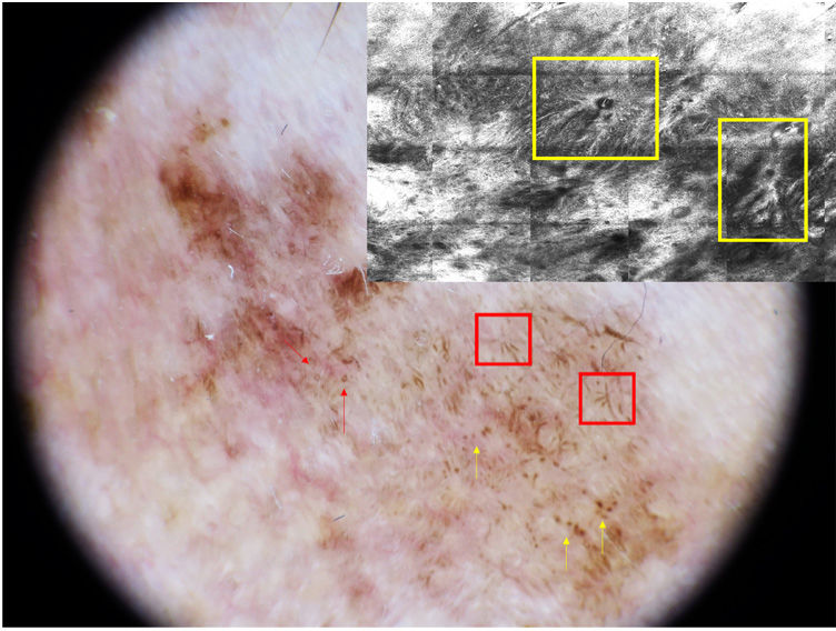
Confocal microscopy revealed abundant perifollicular dendritic cells in the epidermis and atypical junctional thickening arranged around the follicle at the dermal–epidermal junction (Fig. 1, yellow rectangles).
Lentiginous melanoma associated with chronic sun damage may present perifollicular radial lines and blurring of the reticle in dermoscopy as an incipient sign of melanoma (especially when not located on the face).
Perifollicular radial lines in dermoscopy and medusa head-like structures in confocal microscopy play an essential role in the diagnosis of this type of melanoma.


