


Folliculocystic and collagen hamartoma associated with tuberous sclerosis complex was first described by Torrelo et al. in 2012.
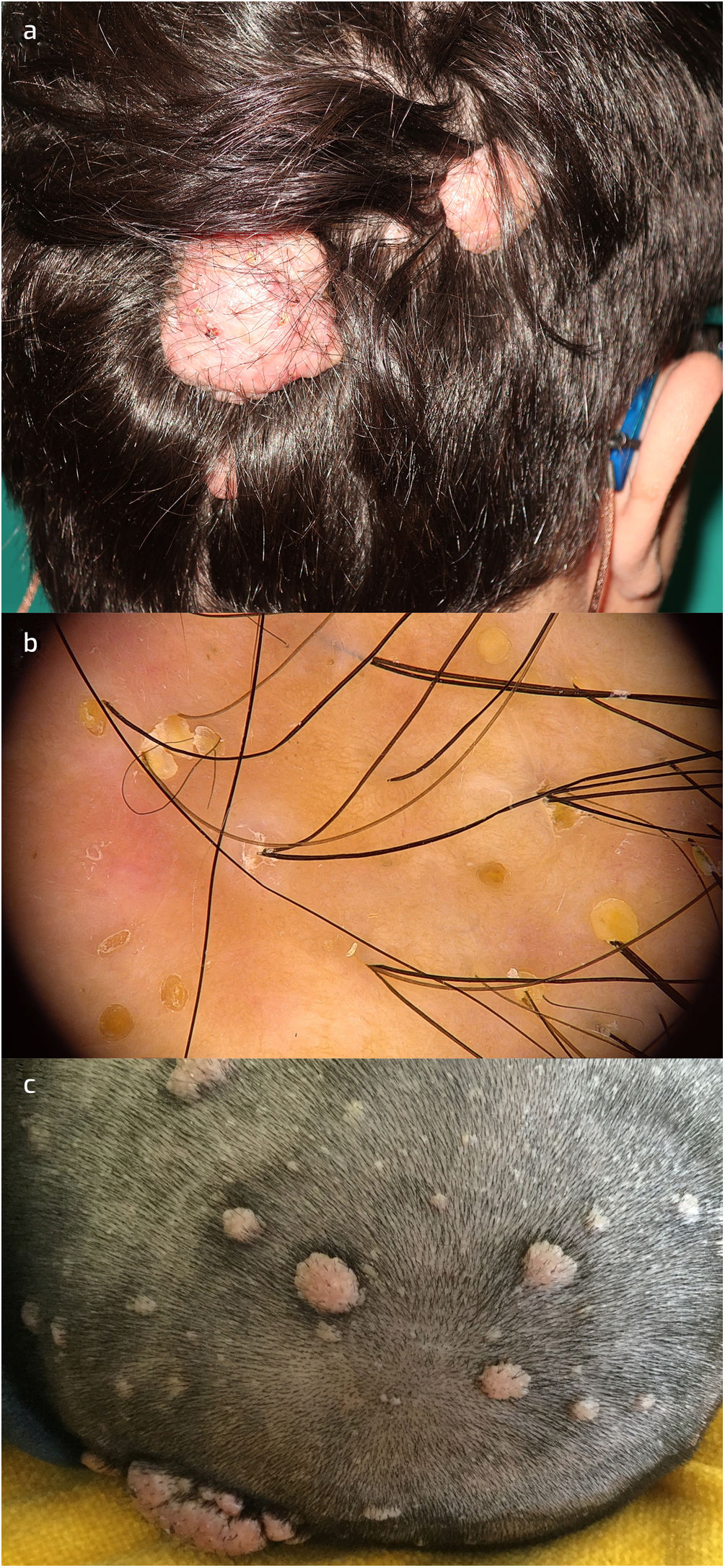
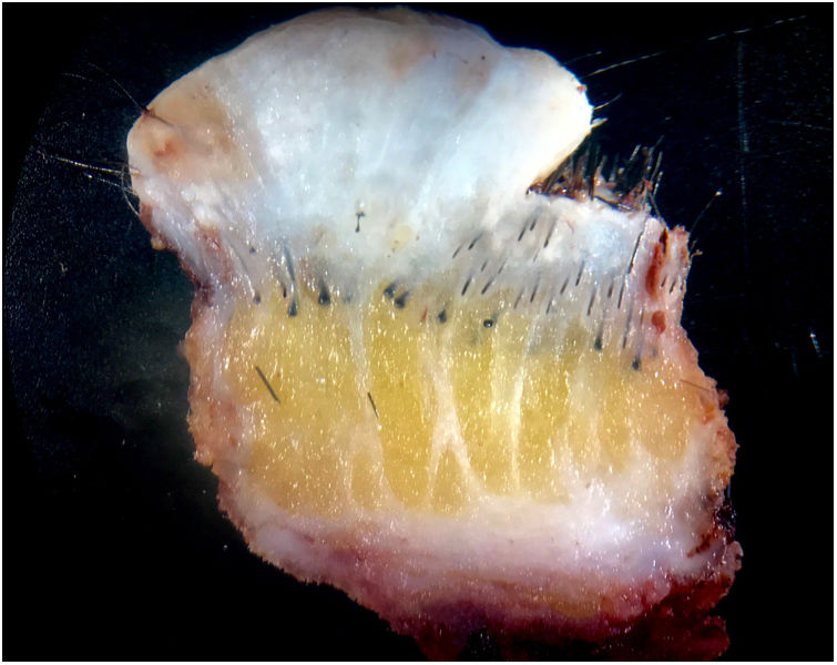
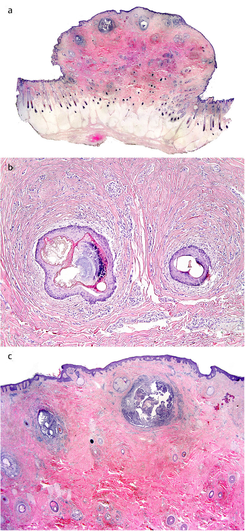
A man in his 20s with the background of tuberous sclerosis complex (TSC) with a “de novo” mutation in TSC2 c.2540t>g (P.l847r). Clinical findings included over three hypopigmented macules, facial angiofibromas, periungual fibromas, cephalic plaque, lumbar shagreen patch, and dental pits. He also had epilepsy and severe mental impairment. Physical examination revealed a polylobulated plaque with soft consistency on the occipital scalp (Fig. 1a), with follicular comedo-like openings and areas of alopecia and clumps of hairs (Fig. 1b). The lesion had been present since birth and grown slowly over time. No history of pain, secretion, or bleeding was reported. An MRI showed cortical and subependymal nodules, but no astrocytomas were detected. Surprisingly, the day before surgery multiple similar, smaller tumors were found all over the scalp after his head had been shaved (Fig. 1c). The largest mass and others of smaller size were completely excised (Fig. 2). Pathology of the specimen revealed a large dermal based exophytic lesion composed of thick collagen bundles in a haphazard distribution (Fig. 3a), which were also arranged concentrically around hair follicles (Fig. 3b). These thickened collagen bundles occupied the entire dermis and reached the superficial subcutaneous septa. There were terminal hair follicles trapped inside the lesion, some of which were cystically dilated, with signs of rupture: suppurative inflammation, foreign-body granulomata, and free hair shafts inside the dermis (Fig. 3c). An infiltrate composed of lymphocytes, plasma cells, and a few eosinophils was present around the ruptured cysts.
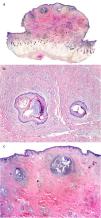
a. Folliculocystic and collagen hamartoma in a patient with tuberous sclerosis complex.
b. Dermoscopy showed yellow plugs in follicular openings, perifollicular desquamation, dystrophic hairs, empty follicular openings, erythema, and some peripilar casts.
c. Multiple hamartomas in the scalp of the same patient.
FCCH was first described by Torrelo et al. in a case series of six male patients with plaques in the abdomen, thigh, back, and scalp. All lesions were solitary, with elastic consistency, irregular surface, and had follicular comedo-like openings and infundibular cysts which occasionally discharged a purulent material.1 The lesions were first noticed at birth or during early infancy. Histopathology showed a diffuse and fibrotic appearance, with septa extending into the subcutaneous tissue and surrounding hair follicles. Eccrine glands and some vessels were involved. Comedo-like openings and cysts containing keratin were described in the dermis.1
Since infundibular cyst formation is not observed in other collagen nevi such as shagreen patches, these features are recognized as a distinctive hamartoma.
After the first report, 7 additional cases have been reported, including the current case. FCCH has been found mostly in males (10 out of 14 cases) and located in the scalp (7 of 13 cases).2–5 A case of a child with TSC and two FCCH in the abdominal wall has been reported.5 To the best of our knowledge this is the first reported case of multiple scalp FCCH.
The diagnosis of multiple lesions went unnoticed during our evaluations at the outpatient clinic and should be actively sought in patients with TSC, and even more so in those with FCCH.
Conflict of interestThe authors declare not to have any conflict of interest.
To Dr. José Luis Rodríguez Peralto and Dr. Luis Javier del Pozo Hernando for their collaboration during the elaboration of the manuscript


