A 48-year-old woman with type 2 diabetes mellitus presented with a >2-year history asymptomatic tumor on the dorsolateral region of her right foot. Physical examination revealed a 1.5cm in diameter exophytic, pedunculated, skin-colored lesion with erythematous areas and verrucous zones (Fig. 1).
Polarized light dermoscopy revealed reddish areas resembling a vascular bed, branched vessels with rounded ends, yellowish structureless areas, and intertwined white areas encircling the vessels (Fig. 2).
Polarized light dermoscopic images of the lower region of the lesion. Red vascular bed-like areas (white circle), branched vessels with rounded ends (black circles), yellowish structureless areas (blue circle), and intertwined white areas surrounding the vessels (black arrow) are observed.
What is your diagnosis?
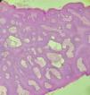
DiagnosisDermoscopic diagnosis suggested eccrine poroma, and histological analysis confirmed this suspicion. The histology showed an epidermis with focal hyperkeratosis and anastomosing cords of cuboidal cells with hyperchromatic, basaloid-appearing nuclei connected by intercellular bridges. These cords were located in the lower third of the epidermis toward the dermis (Fig. 3).
CommentaryEccrine poroma is a rare benign adnexal tumor. It typically presents in older adults without gender preference. Clinically, it appears as a solitary papule, plaque, tumor, or nodule with a smooth, scaly, or verrucous surface. Ulcerations and erosions can occur secondary to friction. Although most lesions are pink, they may also appear reddish, blue, or even black when pigmented.1 They are usually located in acral regions, particularly on palms and soles, due to the high density of eccrine glands, though they can also occur in other areas.2
Dermoscopy is a valuable diagnostic tool. The hallmark findings suggestive of eccrine poroma include branched vessels with rounded ends—formerly called “chalice-shaped” or “cherry blossom” vessels—and intertwined white areas surrounding the vessels. Yellowish structureless areas and reddish vascular bed-like areas are also characteristic,3,4 all of which were present in our patient.
Less common dermoscopic findings include irregular linear vessels, glomerular vessels, bright white areas, atypical reticulation, hemorrhagic crusts, brown structureless areas, keratin, white circles, and regression structures.3,4
In a multicenter study by Marchetti et al.,4 4 dermoscopic patterns were identified: Pattern #1: Most common on hands and feet, showing reddish vascular bed-like areas, branched vessels with rounded ends, and yellowish structureless areas. This pattern was observed in our patient. Pattern #2: Found on the trunk and extremities, with polymorphic vessels, intertwined white areas around vessels, and branched vessels with rounded ends. Pattern #3: Characterized by small lesions anywhere on the body, often without vessels, clinically mimicking basal cell carcinoma. Pattern #4: Found anywhere on the body, featuring large lesions, sometimes pigmented, with blood spots, keratin, and atypical forked vessels.
Eccrine poroma is considered a great mimic, and its primary clinical differential diagnoses include amelanotic melanoma, squamous cell carcinoma, and basal cell carcinoma. Other differentials include pyogenic granuloma, hemangioma, seborrheic keratosis, wart, and melanocytic nevus.1
Treatment of choice is surgical excision. Complete removal is necessary, as a small percentage of lesions may progress to porocarcinom.5
FundingNone declared.


