Información de la revista
Vol. 114. Núm. 4.
Páginas 347 (Abril 2023)
Compartir
Compartir
Descargar PDF
Más opciones de artículo
Vol. 114. Núm. 4.
Páginas 347 (Abril 2023)
Images in Dermatology
Acceso a texto completo
Earlobe Infiltration: A Curious Case of Leukemia Cutis
Infiltración en el lóbulo: un caso curioso de cutis leucémico
Visitas
2271
C. Shajila, D. Sathishkumara,
Autor para correspondencia
dharshsathish@gmail.com

Corresponding author.
, R. Babub, F.N. Aboobackerc
a Department of Dermatology, Venereology and Leprology, Christian Medical College and Hospital, Vellore, Tamil Nadu, India
b Department of Pathology, Christian Medical College and Hospital, Vellore, Tamil Nadu, India
c Department of Clinical Haematology, Christian Medical College and Hospital, Vellore, Tamil Nadu, India
Contenido relaccionado
Actas Dermosifiliogr. 2023;114:T34710.1016/j.ad.2022.02.036
C. Shajil, D. Sathishkumar, R. Babu, F.N. Aboobacker
Este artículo ha recibido
Información del artículo
Texto completo
Bibliografía
Descargar PDF
Estadísticas
Figuras (2)
Texto completo

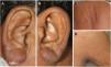
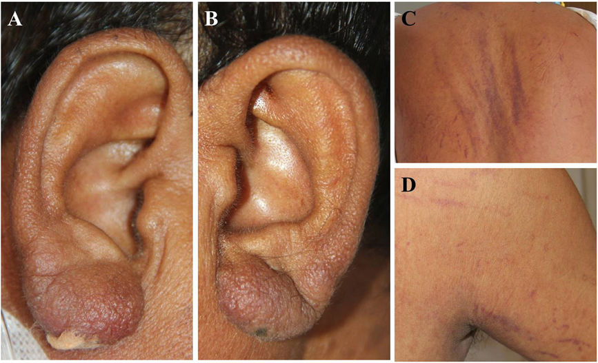
A 45-year-old male presented with intermittent fever, loss of weight and appetite for 2 months. On examination he had generalised lymphadenopathy, faint erythematous infiltration of the earlobes (Fig. 1A and B), and non-palpable, linear purpuras (vibices) involving the trunk and extremities (Fig. 1C and D).

Figure 1
(0,09MB).

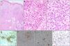
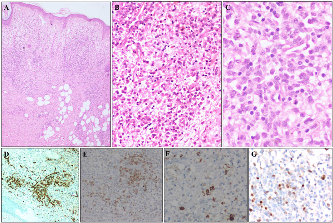
Slit skin smears were negative for acid-fast bacilli. Biopsy from right earlobe revealed moderate-dense dermal infiltrate of atypical lymphocytes displaying high nuclear-to-cytoplasmic ratio (Fig. 2A–C), positive for CD34 (Fig. 2D), CD7 (Fig. 2E), myeloperoxidase (Fig. 2F), TdT (Fig. 2G), consistent with leukemia cutis (LC). Blast cells were detected in peripheral smear (18%) and bone marrow aspirate (85%) and immunophenotyping was consistent with early T-cell precursor acute lymphoblastic leukemia (ALL). Extramedullary involvement was absent and he was initiated on adult ALL protocol.

Figure 2
(0,26MB).

The cutaneous infiltration of leukemic cells in systemic leukemia or LC may manifest as solitary/multiple, localised/disseminated papules, nodules, and plaques, with rare reports of other morphological presentations, often mimicking non-specific skin lesions of leukemia. It is more common among elderly, males, with acute myeloid leukemia or chronic lymphocytic leukemia (CLL).1

Earlobe infiltration is an intriguing, localised presentation of LC, previously reported in CLL, but not in ALL.2 Biopsy was crucial in excluding other clinical differentials like leprosy, lymphomas, pseudolymphomas, granulomatous rosacea, lupus vulgaris, sarcoidosis, chilblain lupus.

References
[1]
G. Wagner, K. Fenchel, W. Back, A. Schulz, M.M. Sachse.
Leukemia cutis – epidemiology, clinical presentation, and differential diagnoses: leukemia cutis.
JDDG J Dtsch Dermatol Ges, 10 (2012), pp. 27-36
[2]
A. Raufi, M. Alsharedi, Y. Khelfa, D.C. Griswold, Y. Lebowicz.
Leukemia cutis in a patient with chronic lymphocytic leukemia presenting as bilateral helical nodules.
SAGE Open Med Case Rep, 4 (2016),
Copyright © 2022. AEDV
Idiomas
Actas Dermo-Sifiliográficas
Opciones de artículo
Herramientas
es en

¿Es usted profesional sanitario apto para prescribir o dispensar medicamentos?

Are you a health professional able to prescribe or dispense drugs?