


An 82-year-old male, Fitzpatrick phototype III, presented to the Dermatology outpatient clinic for a 3-month evolution of an asymptomatic purple nodule on his left supraorbital region. Physical examination disclosed a 0.9cm×1cm nodular, well-defined, purple tumor (Fig. 1).
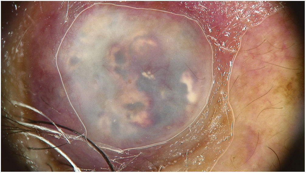
Dermoscopy of the lesion (Fig. 2)
What is your diagnosis?
CommentDermoscopic evaluation revealed white/yellow homogenous areas on a blue-violaceous background, as well as linear irregular vessels. Histopathological exam disclosed the presence of solid nests of basaloid cells with abrupt trichilemmal-type keratinization and ghost cells, confirming the diagnosis of pilomatrixoma.
Pilomatrixoma is a benign soft tissue tumor that originates from the follicular matrix of hair and is also known as Malherbe's calcified epithelioma, due to its tendency toward calcification.1 It usually presents as a single, solid, deep subcutaneous or dermal mass, often on the head or neck.2 There are two incidence peaks, the first in children and adolescents, and a second smaller peak, in older individuals, usually 50–60 years-old.3 Skin lesions are often blue or red in color.1,3 Due to the wide variety of possible clinical findings, clinical misdiagnosis is frequent.1 Common histological features include basaloid cell, calcifications, and ghost cells. Histological subtypes include giant (>5cm in diameter), anetodermic, proliferating and perforating pilomatrixomas.4
The most frequent dermoscopic findings are the white-yellow homogenous areas, irregularly shaped and distributed, that on histology correspond to calcification; white streaks; reddish homogenous areas and vessels, most often hairpin or linear and irregular.2 Additional findings include ulceration, dotted vessels, and structureless blue-gray areas. Specific dermoscopic criteria for melanocytic or nonmelanocytic tumors are absent.2 Although the presence of the criteria mentioned above may suggest the diagnosis of pilomatrixoma, histopathological exam remains essential for confirmation.1
Conflict of interestThe authors declare that they have no conflict of interest.


