


In the existing literature there are few reports describing the development of malignant tumor lesions on port-wine stain capillary malformations. Ultrasound is a novel and informative additional test in patients with these types of skin lesions.
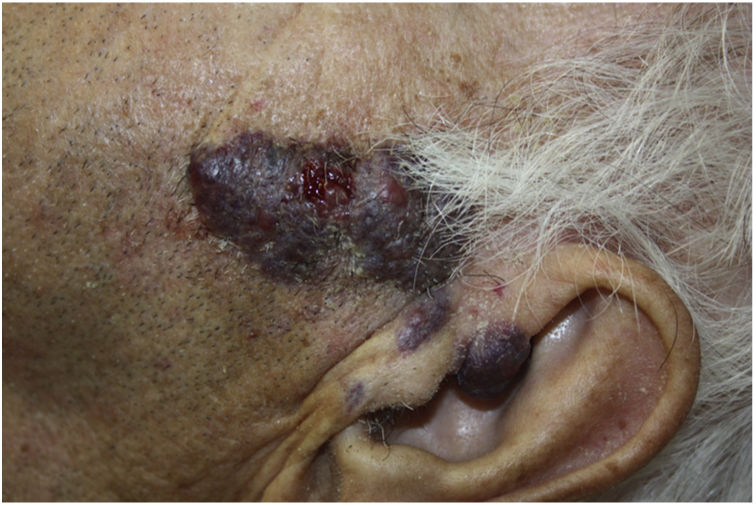
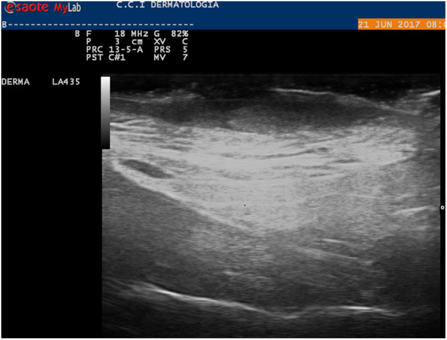
The patient was an 82-year-old man with a history of capillary malformations in the left preauricular region for which he had never been treated. He visited our department after the lesion began to bleed, resulting in the formation of a crust over the central area of the lesion. Physical examination revealed a reddish-purple tumor of approximately 5 cm with a vascular appearance. The central area of the lesion consisted of an erythematous, friable tumor of about 1 cm (Fig. 1). Skin ultrasound revealed an oval hypoechoic lesion of 47 × 4.7 mm in the superficial dermis, and an oval anechoic lesion of 8.3 × 2.2 mm on the skin surface that resulted in erasure of the epidermis with no increase in vascularization (Fig. 2). The tumor was excised respecting the margins observed on ultrasound. Histology confirmed that the lesion was a moderately differentiated squamous cell carcinoma excised with free borders (Fig. 3).

We wish to highlight the utility of skin ultrasound, which can provide important information about lesion location, size, and vascularization, as well as the relationship between the lesion and adjacent major anatomical structures.
Please cite this article as: Oscoz-Jaime S, Azcona-Rodríguez M, Yanguas-Bayona JI. Carcinoma epidermoide sobre mancha en vino de Oporto. Actas Dermo-Sifiliográficas. 2019;110:e1–e2.


