


Photodynamic therapy involves the topical application of a photosensitizer to a lesion, which is then subsequently exposed to a light source. It is mainly used in the nonsurgical treatment of nonmelanoma skin cancer, in which it achieves good response and an excellent cosmetic result. In the last 10 years, photodynamic therapy has also been used with some success in the treatment of plaque-stage mycosis fungoides and has emerged as an alternative to skin-directed therapies. Its main advantages are the good response to treatment, lack of toxicity, and excellent cosmetic results. This article reviews the literature and the practical application of photodynamic therapy in mycosis fungoides.
La terapia fotodinámica (TFD) consiste en la aplicación de un fotosensibilizante tópico en la lesión a tratar y su posterior iluminación con una fuente de luz. Su principal indicación es el tratamiento del cáncer de piel no melanoma sin cirugía con excelente respuesta y resultado cosmético. Su aplicación en las placas de micosis fungoide (MF) en esta última década también ha sido realizada con éxito y se muestra como una alternativa en las terapias dirigidas a la piel. Sus principales ventajas son una buena respuesta, su inocuidad y excelente cosmética. Este artículo revisa los trabajos publicados y la aplicación práctica de la TFD en la MF.
Mycosis fungoides (MF) is the most common primary cutaneous lymphoma and accounts for approximately 50% of these tumors.1 Histologically, MF is characterized by an epidermotropic infiltrate of atypical lymphocytes with cerebriform nuclei.2 Based on clinical presentation, MF is typically classified as patches, plaques, or tumors, though there is actually a wide variety of presentations, all of which are included as variants and subtypes in the latest classification system (2005) of the World Health Organization-European Organization for Research and Treatment of Cancer (EORTC).1
The etiology and pathogenesis of MF are not fully understood, and it has not yet been firmly established whether the T lymphocytes that form the characteristic infiltrate of MF are responding to autoantigens and that their proliferation leads to the formation of a lymphoma or the lesion is a de novo neoplastic proliferation.4 The accumulation of lymphocytes in the skin is thought to be due to a failure of apoptosis rather than increased proliferation.5
MF typically presents clinically as persistent patches or plaques that are sometimes pruritic and that usually affect areas of the skin not exposed to sunlight. These lesions can often remain stable for years or even throughout the patient's lifetime, but progression to disseminated lesions, tumors, or extracutaneous disease can occur, worsening the prognosis and requiring systemic treatment.
Photodynamic therapy (PDT) is a technically simple, local treatment that consists of the application of a topical photosensitizer, occlusion while the photosensitizer is taken up by the target cells, and illumination with an appropriate light source to cause the destruction of those cells. The topical photosensitizers most widely used in dermatology are δ-aminolevulinic acid (ALA) and its ester, methyl-aminolevulinic acid (MAL). Both are precursors of protoporphyrin IX (PpIX), the photosensitive compound, which is produced via the heme synthesis pathway. The main application of PDT is for the nonsurgical treatment of nonmelanoma skin cancer; however, new indications have been developed in recent years and plaque MF is probably the most promising of these.
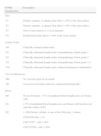
General Therapeutic Approach to Mycosis FungoidesThe staging described in the latest consensus statement published by the International Society for Cutaneous Lymphomas (ISCL)-EORTC6 in 2011 is summarized in Table 1; this statement updates the same group's 2007 staging system3 and the WHO-EORTC system of 2005.1
International Society for Cutaneous Lymphomas–European Organization for Research and Treatment of Cancer 2011 Staging System for Mycosis Fungoides.
| TNMB Classification | Description |
| Skin | |
| T1 | Patches, papules, or plaques that affect < 10% of the skin surface |
| T2 | Patches, papules, or plaques that affect > 10% of the skin surface |
| T3 | One or more tumors ≥ 1cm in diameter |
| T4 | Erythroderma that affects > 80% of the body surface |
| Lymph Nodes | |
| N0 | Clinically normal lymph nodes |
| N1 | Clinically abnormal lymph nodes; histopathology Dutch grade 1 |
| N2 | Clinically abnormal lymph nodes; histopathology Dutch grade 2 |
| N3 | Clinically abnormal lymph nodes; histopathology Dutch grade 3-4 |
| NX | Clinically abnormal lymph nodes without histological confirmation |
| Visceral Metastases | |
| M0 | No visceral organ involvement |
| M1 | Visceral involvement (must be confirmed histologically) |
| Blood | |
| B0 | No involvement: < 5% of peripheral blood lymphocytes are Sézary cells |
| B1 | > 5% of peripheral blood lymphocytes are Sézary cells but does not meet the criteria of B2 |
| B2 | > 1000 Sézary cells/μL or one of the following 3 criteria: |
| CD4/CD8 ratio > 10 | |
| CD4+/CD7− cells > 40% | |
| CD4+/CD26− cells > 30% | |
| Stage | TNMB Classification |
| IA | T1, N0, M0, B0-1 |
| IB | T2, N0, M0, B0-1 |
| IIA | T1-2, N1-2-X, M0, B0-1 |
| IIB | T3, N0-1-2-X, M0, B0 |
| IIIA | T4, N0-1-2-X, M0, B0 |
| IIIB | T4, N0-1-2-X, M0, B1 |
| IVA1 | T1-4, N0-1-2-X, M0, B2 |
| IVA2 | T1-4, N3, M0, B0-2 |
| IVB | T1-4, N1-2-3-X, M1, B0-2 |
Abbreviation: TNMB, tumor, node, metastasis, blood.
Source: Olsen et al.6
The variability in the clinical presentation and clinical course of MF has led to the appearance of numerous therapeutic options and complex algorithms. A number of guidelines on the treatment of MF and Sézary syndrome have been published, including those of the European Society of Medical Oncology7 and of the National Cancer Center Network.8 It is important to realize that there have been very few clinical trials in MF, as it is a rare dermatosis, and the evidence on which these guidelines are based is therefore limited.
For the management of early MF (stages IA, IIA, IIB) the guidelines propose 2 forms of skin-directed therapy, one for limited or localized skin disease and another for generalized or widespread involvement.3,6–8 These options are summarized in Table 2. Topical corticosteroids are the most widely used skin-directed therapy in early MF. If they are not effective, the common alternatives are nitrogen mustard, carmustine, topical bexarotene, local radiotherapy, surgical excision, and topical imiquimod. Phototherapy (narrow-band UV-B or psoralen–UV-A), alone or in combination with systemic therapy (bexarotene, interferon, or methotrexate), is an ideal treatment when plaques are widespread.
Recommended Treatments for Early Mycosis Fungoides.
| Localized disease | Widespread disease |
| Topical corticosteroids | Topical corticosteroids |
| Topical chemotherapy (nitrogen mustard, carmustine) | Topical chemotherapy (nitrogen mustard, carmustine) |
| Local radiotherapy | Phototherapy: UV-B, psoralen–UV-A |
| Topical retinoids (bexarotene, tazarotene) | Total skin electron beam radiation |
| Phototherapy: UV-B, psoralen–UV-A | |
| Topical imiquimod |
Because PDT is applied locally and has limited penetration into the skin, it has a promising role as a one of the localized skin-directed treatment options. In advanced stages of MF (IIB or higher), PDT is of no interest precisely because it is a localized treatment9; however, it may occasionally be used as palliative treatment for individual plaques or tumors.
Mechanism of Action of PDTIn 1994, Svanberg et al.10 published good results after using PDT to treat 2 patients with plaque-stage MF. The same year, Boenhcke et al.11 demonstrated how PDT inhibited lymphocyte proliferation in MF plaques, both in vivo and in vitro. Later, in 1995, Rittenhouse-Diakun et al.12 showed that malignant lymphocytes in MF lesions had higher expression of CD71 (the transferrin receptor) than normal lymphocytes, and that this receptor gave the cells a greater capacity to absorb iron and to accumulate PpIX, making them more susceptible to treatment with PDT. Edstrom et al.,13 in 2000, demonstrated a reduction of the CD71+-lymphocyte infiltrate in MF plaques after treatment with PDT due to decreased cell proliferation. Previously, in 1998, the same authors had demonstrated a decrease in the number of typical CD4+/CD7− lymphocytes in the infiltrate of MF plaques after treatment with PDT, again due to decreased proliferation, though he was unable to demonstrate the implication of apoptotic mechanisms.14 All these findings support the ability of PDT to selectively destroy the malignant lymphocytes in MF lesions.
The greater capacity of malignant lymphocytes to accumulate PpIX intracellularly appears to be related to their intrinsic ability to produce and retain this compound due to slower metabolism of the heme group. Accumulation is enhanced because of certain metabolic changes: an increased activity of the rate-limiting enzyme in the production of the heme group (porphobilinogen deaminase)15; acceleration of the cell cycle in the tumor cells, which increases their capacity to take up ALA; and a higher density of mitochondria and a lower intracellular pH.16 In addition, the changes in the stratum corneum of the epidermis in the plaques favor penetration of the photosensitizer into the skin.17
The histological response to treatment has been evaluated in a number of studies, though the results have been variable. Ammann et al.18 and Díez-Recio et al.19 reported complete histological cure and an absence of infiltrates of atypical lymphocytes in the plaques showing complete clinical response to treatment. Both groups described pigmentary changes with the presence of melanophages, dermal fibrosis, epidermal atrophy, and residual lymphocytes in the infiltrate. Edstrom et al,13 on the other hand, detected a residual infiltrate of atypical lymphocytes in some cases in lesions showing complete clinical response. These findings could be explained by the depth of light penetration, which might not be sufficient to treat the deepest lymphocytes in the MF plaques. Eich et al.20 treated 8 patients with MF tumors; the subsequent biopsy showed resolution of the infiltrate down to a depth of 1.5mm and the continued presence of atypical lymphocytes in deeper tissue. These histological findings mean that patients with a complete response must be followed for possible recurrence, as published studies have not clearly established that PDT has the ability to completely eliminate the atypical lymphocytes in the plaques. However, the practical repercussions of this finding are minimal, as patients with plaque MF must always return for periodic check-ups.3,6–8
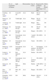
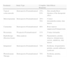
Clinical StudiesThe published studies of the use of PDT in MF are summarized in Table 3.10,13,14,21–28 Those studies included a total of 45 patients with 75 MF lesions treated with PDT. The majority of authors have worked with ALA; the ester, MAL, was only used in 2 studies. Red light (coherent or noncoherent), which offers greater penetration into the skin, was employed most often. The overall response rate of the MF lesions treated in all these small case series was 84% (78% in patches and 84% in plaques). Although more heavily infiltrated lesions will, in theory, have a poorer response to this treatment, remission was observed in all the tumor lesions described in these reports. Publication bias could account for these observations, as successful treatments are more likely to be published than failures. A review comparing PDT to other conventional treatment options for plaque-stage MF (Table 4)29 found that response rates were similar or even slightly better with PDT, which had the added advantage of fewer side effects. However, this review provided only an approximate assessment of relative merits, as the studies compared were not homogeneous, used different methods, and, above all, were not clinical trials. PDT is not comparable to phototherapy (which therefore does not figure in Table 3) as PDT is a local treatment that is mainly indicated in patients with a small number of lesions (maximum of 3 or 4), whereas phototherapy is particularly useful in widespread lesions.
Summary of Published Studies on the Treatment of Mycosis Fungoides With Photodynamic Therapy.
| No. of Lesions/No. of Patients | Light | Photosensitizer | Type of Lesion | Responses/No. of Patients Treated (Percentage of CRs) | Follow-up Period, mo | |
| Svanberg et al., 199410 | 4/2 | Laser,630nm | ALA | Patch | CR 1/2 (50%) | NS |
| Wolf et al., 199421 | 2/2 | Visible light | ALA | Plaque | CR 2/2 (100%) | 3–6 |
| Amman et al., 199518 | 1/1 | Visible light | ALA | Plaque | CR 1 (100%) | NS |
| Edstrom et al., 199814 | 5/1 | Red light, 630nm | ALA | NS | CR 4/5 (80%) | NS |
| Wang et al., 199922 | 3/1 | Red light, 635nm | ALA | Plaques | CR (100%) | 33 |
| Orenstein et al., 200923 | 6/2 | Noncoherent light,580–720nm | ALA | 1 patch2 tumors | CR (100%) | 24 |
| Markham et al., 200124 | 1/1 | Noncoherent light,580–740nm | ALA | Tumor | CR (100%) | 12 |
| Edstrom et al., 200113 | 12/10 | Noncoherent light,600–730nm | ALA | 10 patches2 tumors | 9/10 plaques CR (90%)2/2 tumors IR | 4–19 |
| Leman et al., 200225 | 2/1 | Red light,630nm | ALA | Patches | CR (100%) | 12 |
| Coors et al., 200426 | 7/5 | Noncoherent light, 60–160nm | ALA | 5 patches2 tumors | CR (100%) | 12–18 |
| Zane et al., 200627 | 5/5 | Red light,635nm | MAL | Patches | 4 CR (80%)1 PR | 12–34 |
| Díez-Recio et al., 200719 | 2/2 | PDL, 585nm | ALA | Plaques | CR (100%) | 34 |
| Fernández-Guarino et al., 201029 | 24/12 | 630 | MAL | Plaques | CR 6/12 (50%) | 6–36 |
Abbreviations: ALA, δ-aminolevulinic acid; CR, complete response; IR, incomplete response; MAL, methyl-aminolevulinic acid; NS, not specified; PDL, pulsed dye laser; PDT, photodynamic therapy; PR, partial response.
Reported Efficacy and Side Effects of the Standard Treatments for Mycosis Fungoides Plaques and a Comparison with Photodynamic Therapy.
| Treatment | Study Type | Complete Response | Side Effects |
| Topical corticosteroids | RetrospectiveNonrandomized | 25%–63% | Skin atrophyShort duration of the therapeutic effect |
| Metoclopramine | RetrospectiveNonrandomized | 26%–76% | Contact dermatitisSecondary skin tumors |
| Carmustine | RetrospectiveNonrandomized | 86% | Bone marrow suppressionTelangiectasia |
| Bexarotene | ProspectiveNonrandomized | 21% | Contact dermatitis |
| Radiotherapy (Electron beam) | Meta-analysis | 96% | Pigmentation, pruritus, alopecia, telangiectasia, xerosis, anhidrosis, skin tumors |
| Imiquimod | RetrospectiveNonrandomized | 70% | Erythema, desquamation, pruritus, pseudo-influenza syndrome |
| Photodynamic Therapy | RetrospectiveNonrandomized | 78% | Erythema, pruritus, blisters |
Source: Whittaker et al.29,30
The majority of the studies have used long wavelengths, around 600nm, in the final absorption band of PpIX, in order to achieve better penetration into the dermis and thus act on the deepest areas of the infiltrate. Both laser and noncoherent light sources have given good results.
Despite the simplicity of the technique, the optimal parameters have not yet been defined. Both ALA and MAL have been found to be effective, and various occlusion times have been used in different protocols. Light dosimetry and the frequency of the sessions have varied considerably between studies, though all described the need for multiple sessions.18,25
Side EffectsThe main advantages of PDT in the treatment of plaque-stage MF are that it is innocuous, noninvasive, convenient, technically simple, and relatively selective, and that it has no carcinogenic potential and gives an excellent cosmetic result.
PDT sessions are usually well tolerated, although patients have described a mild burning sensation in most studies. In the early studies, light was applied at high intensities for short periods of time, but the lower intensities applied in later studies caused less pain while still achieving good results.13 Pain during the illumination appears to be very variable between patients,27 and excellent tolerance was reported in the studies that used MAL rather than ALA as the photosensitizer.27,28
Erythema and edema are common in the treated plaques, but scabs and erosions are rare. When they do appear, they typically resolve in 2 to 3 weeks. Mild pigmentary changes, most commonly hyperpigmentation, or hair loss may develop in lesions with a complete response.27,28
Diagnostic FluorescenceThe fluorescence pattern of MF lesions was described by Orestein et al.23 as weak and diffuse in the patches and as intense with clearly defined borders in the plaques and tumors. Those authors also reported that the intensity of fluorescence decreased more rapidly in patches than in tumors.
In our experience, MF plaques show intense fluorescence with sharp borders.28 The fluorescence decreases as the clinical response develops, and disappears in lesions once a complete response has been achieved. Diagnostic fluorescence therefore appears to be a useful tool for monitoring treatment, though there are no studies to clarify whether it is superior to clinical evaluation.
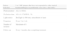
Practical Aspects of ManagementPDT is useful in patients with MF with plaques that are isolated or confined to a limited area. A possible additional indication for PDT is when a good cosmetic result is required, on the face, for example, or in areas at risk of poor healing, where radiation therapy, nitrogen mustard, or carmustine can leave permanent and visible scars. Plaques situated in the axillary or inguinal skin folds that are not accessible to phototherapy can also be treated with PDT (Table 5). Patients with multiple MF plaques are not good candidates for PDT because they require treatment to multiple areas; these patients are better managed using phototherapy.
Practical Aspects of Photodynamic Therapy for the Treatment of Plaque-Stage Mycosis Fungoides.
| Patient Selection | 1 or 2 MF plaques that have not responded to other topical treatmentsPlaques in problematic areas (face, skin folds) |
| Photosensitizer | ALA or MAL |
| Occlusion time | ALA 4–12 h/MAL 3h |
| Light source | Red light (±600 nm), noncoherent or laser |
| Frequency | Every 2 to 4 wk |
| Number of sessions | Maximum of 6 |
| Follow-up | Every 3 months after completing treatment |
Despite the simplicity of applying PDT, the optimal parameters for the occlusion time, illumination time, and fluence have not been established because no clinical trials have been performed. Both ALA and MAL have been used with good results, but the occlusion times have varied from one study to another. In the absence of further studies, the occlusion times for ALA should be from 4 to 16hours and for MAL of 3hours.21–28
The most appropriate light sources provide red light at around 630nm, which achieves deepest penetration and covers the spectrum of PpIX in its final absorption band. Both coherent and noncoherent light sources have proven to be effective, and the choice will depend on system availability or the dermatologist's preference. A summary of the differences between the 2 light sources is given in Table 6. Pulsed dye laser is the most widely used and most suitable laser light source for PDT, as it emits light at 595nm (close to the optimal 630nm).
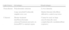
Differences Between Coherent and Noncoherent Light Sources and Their Application in Photodynamic Therapy.
| Light Source | Advantages | Disadvantages |
| Noncoherent | Polychromatic emission | Lower intensity |
| Large areasSafeTechnically simpleLower cost | Marked thermal effectMore difficult to control exact dose | |
| Coherent | Shorter treatment timeMonochromatic emissionTreatment precisely on lesionsPDT to internal organs | Cannot be used on large areasTechnically more complexSpecific installationMore expensive |
Dosimetry varies considerably from one study to another. In general, as there are no studies specifically dealing with MF, we should apply the same doses as are used in approved indications that are routinely treated in order to achieve efficacy without side effects. In the case of laser light sources, despite their effectiveness in many studies, the optimal parameters remain unknown, even for the treatment of approved indications. As the objective is to illuminate the areas to be treated, low (subpurpuric) doses should be applied in several passes until the whole plaque is treated.
Multiple PDT sessions are required to treat MF, although the exact frequency of the sessions also remains undefined. The majority of authors administer sessions every 2 to 4 weeks, a frequency that allows any scabs to resolve before the following session and promotes adherence to treatment, which requires the patient to come to a specialist center. The total number of sessions will depend on the clinical response, and varies considerably between studies (range, 2–18 sessions).21–28 A total of 6 sessions may be considered adequate before interrupting treatment and considering other options if no response is observed.
Tumors should not be treated with PDT. Despite the good results published by some authors, others have reported no reponse.13 It should not be forgotten that red light penetrates 1 to 2mm into the skin, and deeper lymphocytes therefore remain untreated.20 Treatment failure in large plaques has been reported in some studies, and PDT is therefore not recommended for lesions over 7.5cm in diameter.
Patients must always receive periodic check-ups after treatment as clinically resolved plaques may still have histological evidence of disease, and late recurrences have been reported.13,19,20
ConclusionsPDT is a good, well-tolerated option for the treatment of localized MF plaques and provides a good cosmetic result. It is particularly useful in plaques that do not respond to the usual treatments and those located in problematic sites, such as the face or skin folds, or in areas at risk of poor healing. It is not suitable for the treatment of MF tumors or large or very numerous plaques. PDT leads to clinical improvement in MF plaques but is not curative, and periodic follow-up after treatment is therefore necessary.
Ethical DisclosuresProtection of human and animal subjectsThe authors declare that no experiments were performed on humans or animals for this investigation.
Confidentiality of dataThe authors declare that no private patient data are disclosed in this article.
Right to privacy and informed consentThe authors declare that no private patient data are disclosed in this article.
Conflicts of InterestThe authors declare that they have no conflicts of interest.
Please cite this article as: Fernández-Guarino M, Jaén-Olasolo P. Terapia fotodinámica en micosis fungoides. Actas Dermo-sifiliogr. 2013;104:393-9.


