
Tumors with myoepithelial differentiation include mixed tumor, myoepithelioma, and myoepithelial carcinoma. They tend to involve the salivary glands, but they may also be located in the sinonasal area, in the larynx and lungs, and on the skin. Skin tumors with myoepithelial differentiation include chondroid syringoma (mixed tumor), cutaneous myoepithelioma, malignant chondroid syringoma, and myoepithelial carcinoma.1–3 The syncytial variety of cutaneous myoepithelioma has been described in recent years.4
A 33-year-old man with no relevant past personal history visited our department with a papule measuring 0.2 cm in diameter. The papule had appeared 2 years earlier on the right flank and was slightly erythematous and painful. It was not associated with signs of bleeding or growth. A clinical diagnosis of angioleiomyoma and angiolipoma was considered and an excision biopsy of the lesion was performed.
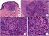
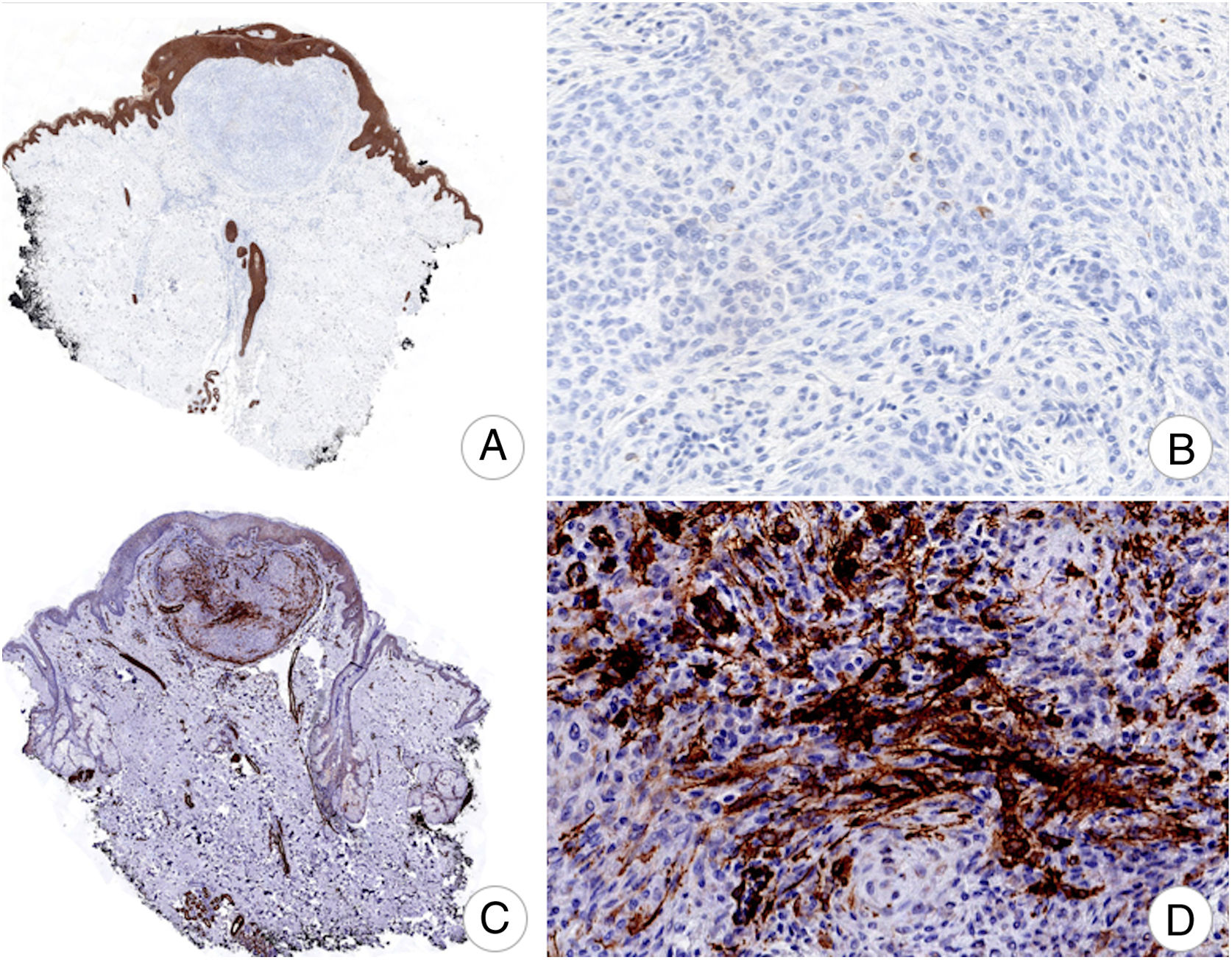
Histology revealed an elevated sessile lesion due to the presence of a solid, well circumscribed, nodular tumor with lobular edges, located on the superficial third of the reticular dermis (Fig. 1A). The lesion consisted of fusiform and epithelioid cells with an oval or rounded nucleus, with no significant atypia, variable quantities of cytoplasm, poorly defined edges, and practically no interstitial component (Figs. 1B-D). A maximum of 4 mitotic figures for every 10 fields at a magnification of 40×. No tumor necrosis was observed.

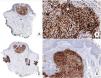
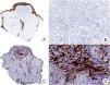
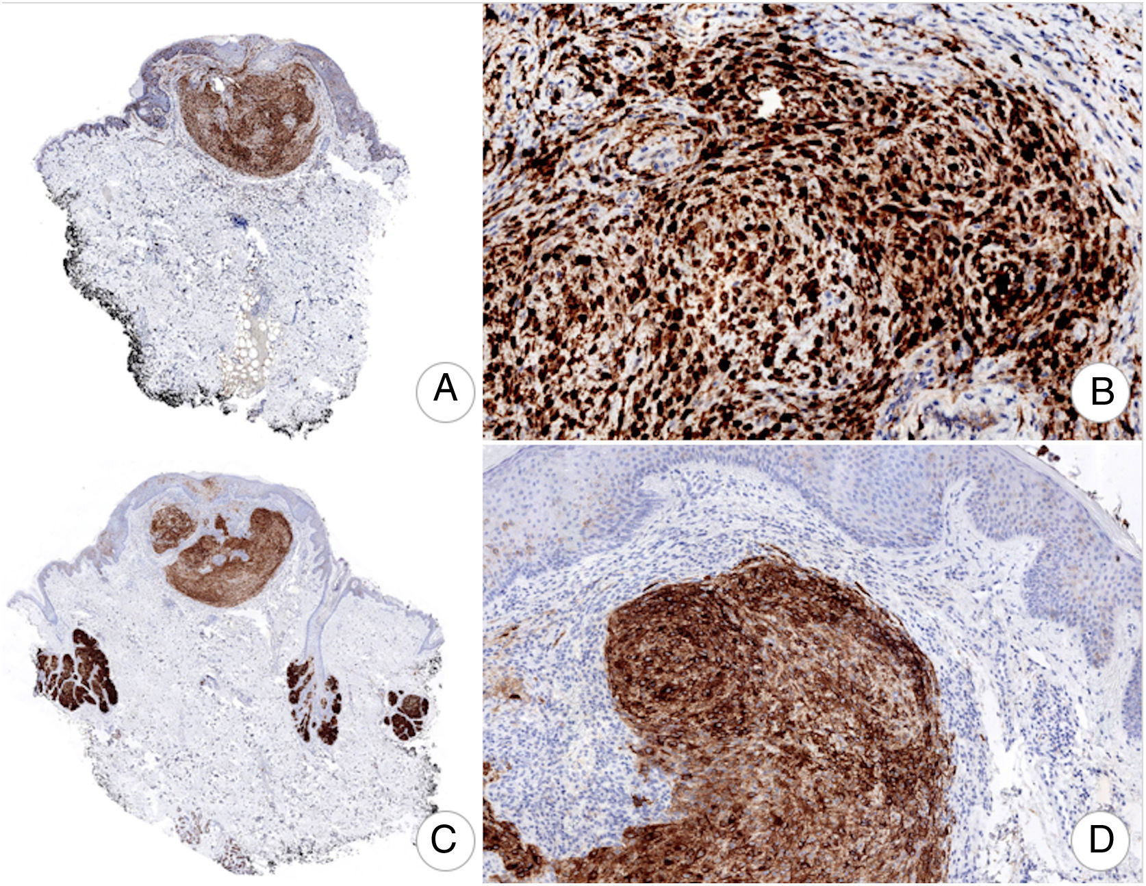
The cancerous population showed diffuse and generalized positive immune staining for S100 and EMA (Fig. 2) and was focal for smooth muscle actin (Figs. 3C and 3D) and caldesmon. No immune staining was observed for cytokeratins AE1-AE3 (Figs. 3A and 3B), Melan-A, desmin, glial fibrillary acidic protein, p63, or claudin-1.
An RT-PCR and EWSR1 rearrangement (EWSR1-POUF5F1, EWSR1-ZNF444 y EWSR1-PBX1) sequencing study was performed5 and the results were negative.
Syncytial myoepithelioma is a rare tumor that presents clinically as a papule or nodule on the limbs of middle-aged men. Histologically, it is a solid tumor with fusiform cells or histiocytes with a pale eosinophilic cytoplasm and vesicular nucleus, with sparse stroma.5,6
It does not usually present mitosis, necrosis or lymphovascular invasion, but in rare cases, up to 4 mitotic figures per 10 fields at 40× have been reported.7 The diagnostic criteria for cutaneous myoepithelial carcinoma are not well established, but tumors with marked cytologic atypia, a high mitotic index, and necrosis show more aggressive behavior, with increased probability of recurrence and distant metastasis.5,7–9
Fifty percent of cutaneous syncytial myoepitheliomas present rearrangement of the EWSR1 gene.4,5,7
Myoepithelial tumors usually express cytokeratins and the S100 protein. Myoepithelioma, however, presents positive immune staining for EMA and S100 protein, and most cases are negative for cytokeratins.5
The differential diagnosis includes epithelioid benign fibrous histiocytoma, juvenile xanthogranuloma, melanocytic lesions, and epithelioid sarcoma.5,9
Epithelioid benign fibrous histiocytoma presents as a dermal nodule of epithelioid cells, frequently with binucleation, positive for EMA, with a fibrovascular stroma. This lesion, however, does not present a syncytial architecture or positive immune staining for S100 protein, or for GFAP or p63, as is the case with syncytial myoepithelioma.4,7,9
In the early stages, juvenile xanthogranuloma presents as an exophytic lesion with eosinophilic histiocytes that does not usually present mononucleated or multinucleated lipidized cells (Touton cells).9 However, it tends to affect children, immune staining is positive for CD163, CD68, and is negative for EMA and S100 proteins.4
Cutaneous syncytial myoepithelioma consists of a combination of epithelioid cells, histiocytes, and fusiform cells. Histology of Spitz nevus shows nests of melanocytic cells with frequent maturation with descent and no syncytial architecture. Both tumors are positive for S100, but positivity for Melan A, HMB-45, and MiTF, and negativity for EMA and GFAP favor a diagnosis of Spitz nevus.4,5,7,9
Epithelioid sarcoma usually presents a combination of epithelioid and fusiform cells with cellular atypia and infiltrative growth with frequent satellite nodules. In some cases, however, it presents with a pattern of mild atypia. Both lesions are positive for EMA, but epithelioid sarcoma is positive for cytokeratins and CD34, negative for myoepithelial markers such as S100 proteins and GFAP, and often presents loss of immune staining for INI-1.5,7,9
The differential diagnosis of a painful skin lesion includes angiolipoma, neuroma, glomic tumor, schwannoma, leiomyoma, eccrine spiradenoma, and dermatofibroma.10
Treatment of cutaneous syncytial myoepithelioma involves wide resection. Our patient presented a good clinical course with no evidence of local recurrence or metastasis in follow-up examinations, in line with the information reported in the literature.
In conclusion, we report a case of painful cutaneous syncytial myoepithelioma. This is a very rare benign skin tumor. Its clinical presentation is nonspecific and diagnosis requires a histopathology examination and a complete immunohistochemistry panel to differentiate it from other lesions associated with a worse outcome. The presence of rearrangement of the EWSR1 gene may aid the diagnosis. Treatment involves wide resection of the lesion, with an excellent prognosis.
Conflicts of InterestThe authors declare that they have no conflicts of interest.
Please cite this article as: Guillen-Climent S, et al. Mioepitelioma sincitial cutáneo doloroso: desde la clínica inespecífica al diagnóstico histopatológico. Actas Dermosifiliogr. 2020;111:173–175.


