


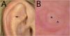
A 72-year-old woman with a history of nonmelanoma skin cancer visited our department for a skin check-up. On inspection, the only finding of note was a lesion on the right antihelix, which had appeared an unknown length of time earlier. The lesion was a slightly erythematous millimetric papule (Fig. 1). No other similar lesions were found on the rest of the skin surface.
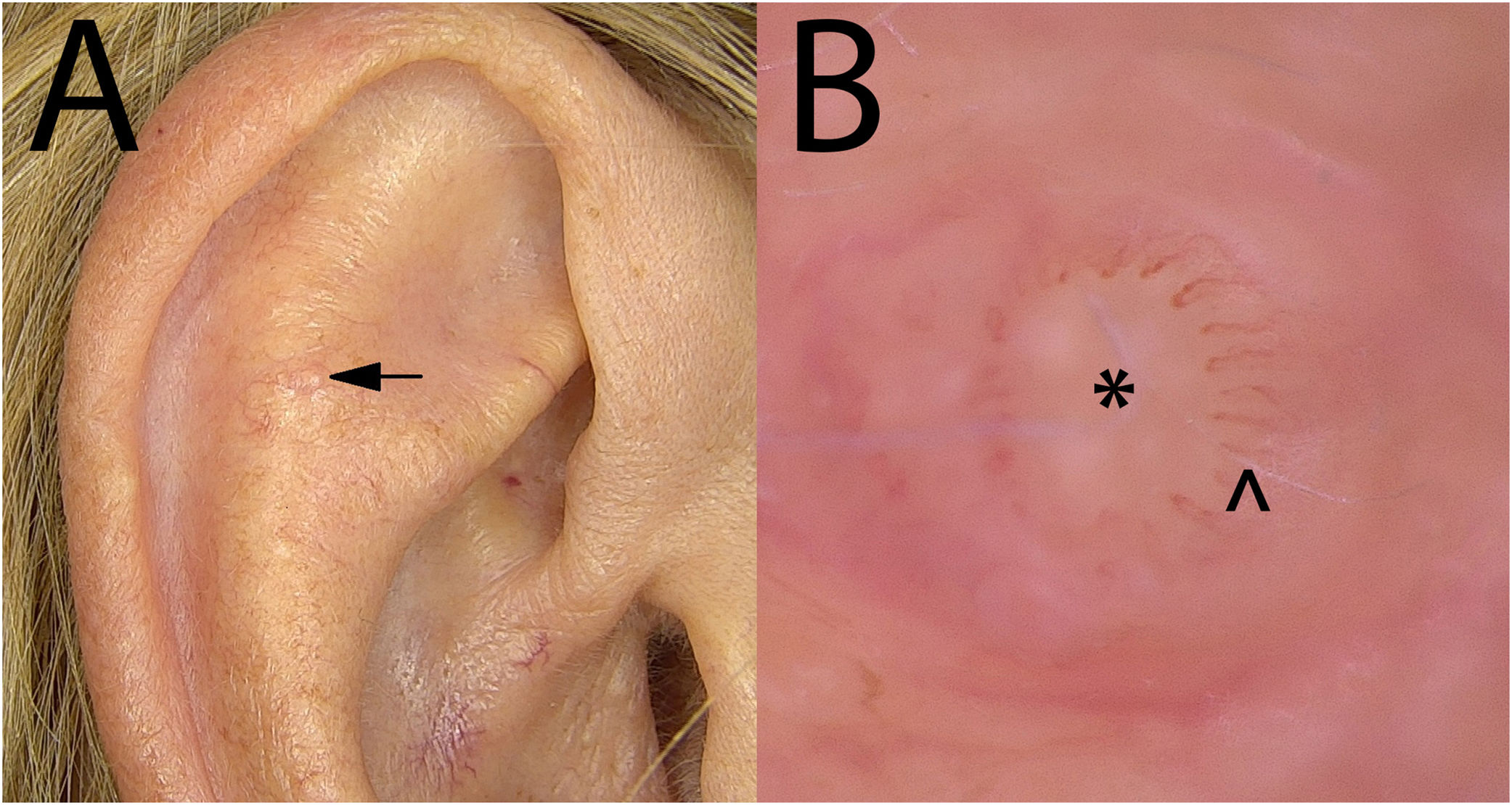
A, Clinical presentation of the lesion on the right antihelix. B, Dermoscopic image of the lesion using contact dermoscopy and 60× magnification (image acquisition was very difficult given the very small size of the lesion). Slightly whiteish central area without structure (*) and radially distributed hairpin vessels (^).
Trichilemmoma or tricholemmoma (Fig. 2).
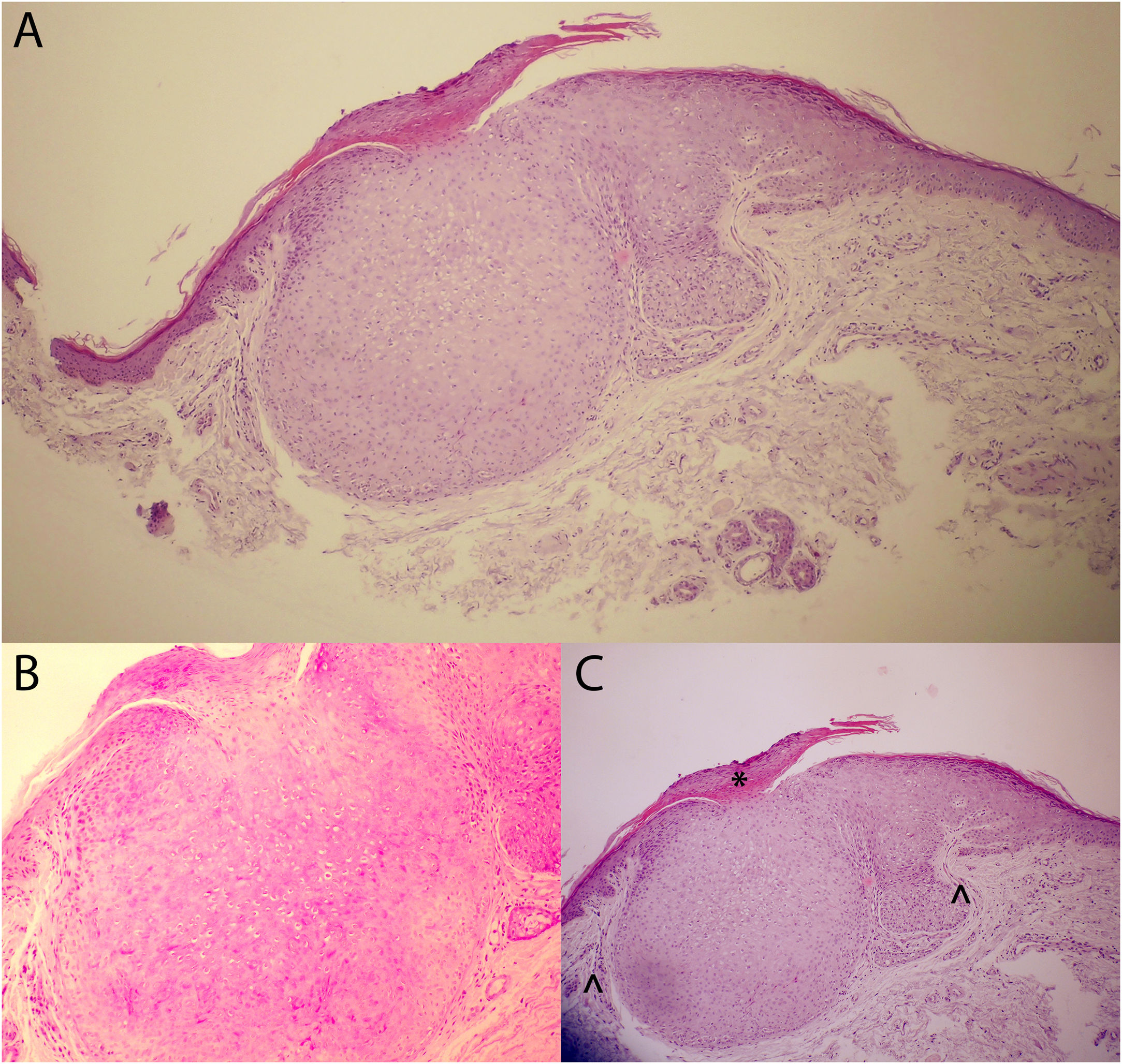
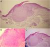
Histopathology findings of the fully excised lesion. Of note is the presence of at least one hyperplasic infundibulum with an epithelium reminiscent of that of the external root sheath of the hair follicle, with hypergranulosis and parakeratosis, consisting of clear monomorphic cells with a pale eosinophilic cytoplasm (due to the presence of glycogen) and a small nucleus. A, General view. B, PAS staining showing the glycogen of the cells that constitute the benign tumor. C, Detailed image with histology-dermoscopy correlation of the parakeratosis (*) and vessels surrounding the lesion (^).
Dermoscopy revealed hairpin vessels in a radial distribution around the periphery of the lesion, with an unstructured whiteish central area with an appearance reminiscent of an ostium. An excision biopsy of the lesion made it possible to reach a diagnosis of trichilemmoma, with some findings that supported the dermoscopic and histologic correlation (Fig. 2).
Trichilemmoma is a benign follicular tumor that is most commonly found on the central facial area, although it may be located on any part of the skin, except glabrous skin. Solitary trichilemmomas are common in adulthood, show no predominance by sex, and are asymptomatic. Different dermoscopic findings have been reported or this entity, including whiteish perivascular halos,1 red iris-like structures, and occasionally, a hyperpigmented halo.2
If it presents in multiple form, a diagnosis of Cowden syndrome should be considered. If it presents in solitary form, as in our case, clinical diagnosis is practically impossible. For this reason, we believe that dermoscopy allows for a more approximate differential diagnosis, even in such incipient lesions as that of our patient. Nevertheless, the differential diagnosis, given the clinical and dermoscopic findings, should be established with other lesions such as an incipient keratoacanthoma or a cystic lesion such as an epidermal inclusion cyst.
Conflicts of interestThe authors declare that they have no conflicts of interest.
Please cite this article as: Martin-Gorgojo A, Bru-Gorraiz FJ, Pascual-Rodríguez E. Pápula milimétrica en antehélix constituida dermatoscópicamente por corona de vasos en horquilla. Actas Dermosifiliogr. 2021;112:837–838.


