
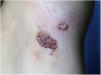
A 15-year-old male presented with asymptomatic, erythematous, grouped vesicles over left axilla noticed since five years of age which did not respond to any topical treatments (Fig. 1).
What is your diagnosis?Diagnosis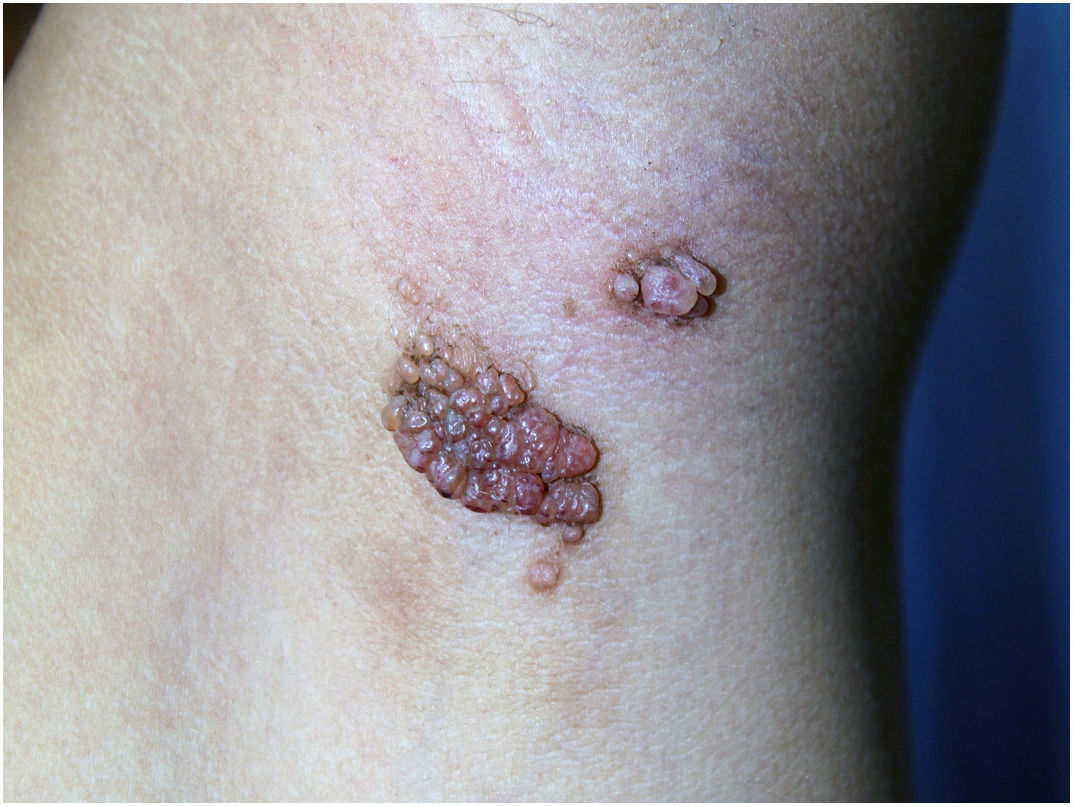
Lymphangioma Circumscriptum.
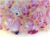
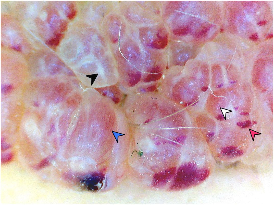
CommentaryPolarized light dermoscopy revealed multiple erythematous lacunae separated by white septa and linear vascular structures (Fig. 2). “Hypopyon sign” which refers to the two-tone lacunae due to the settling of blood in the lowest part of the lacunae was observed (Fig. 2). With the provisional diagnosis of lymphangioma circumscriptum (LS), we performed a biopsy which confirmed the diagnosis.
Cutaneous lymphangiomas are rare vascular tumors divided into two major groups: superficial and deep.1 Lymphangioma circumscriptum, also known as superficial lymphatic malformation is the most common type among these tumors. LS is clinically characterized by clusters of translucent vesicles usually presenting soon after birth. The differential diagnoses include hemangiomas, angiokeratomas, angiosarcomas, lymphangiectasia, hemangiomas, cutaneous metastases, warts and molluscum contagiosum.1–3 Histologically, LS displays many dilated lymphatic channels located in the superficial dermis which may extend to the reticular dermis or subcutaneous tissue.1
Presence of lacunae is the most commonly described histological feature of LS which correspond to the dilated, thin-walled lymphatic vessels in the papillary dermis.4 These lacunae are either filled with lymphatic fluid or red blood cells which gives their characteristic whitish or reddish color.4 Gencoglan et al.5 described “hypopyon sign” occurring due to the precipitation of red blood cells as the differentiating feature between LS and hemangioma. The presence of white lines separating the lacunae has been attributed to the presence of fibroplasia which is seen in many cases of LS.4 Zaballos et al.4 correlated the presence of vascular structures to the micro-shunts between small blood vessels and lymphatic channels.
The presence of lacunae and vascular structures has been described as the most common dermoscopic feature of LS while the “hypopyon sign” has been proposed as the most distinguishing feature of LS.4,5 These dermoscopic features in combination help to distinguish LS from the other conditions included in the differential diagnoses. We present this case for its educational value as all of the previously described dermoscopic patterns of LS can be clearly appreciated.
Conflict of interestThe authors declare that they have no conflict of interest.
We would like to thank Professor Dr. T.S. Rao and Dr. Subechhya Jaiswal for their support during the interpretation of histopathological slides.
Please cite this article as: Mathur M, Acharya P, Karki A. Vesículas rojas agrupadas en la axila. Actas Dermosifiliogr. 2020. https://doi.org/10.1016/j.ad.2019.01.024


