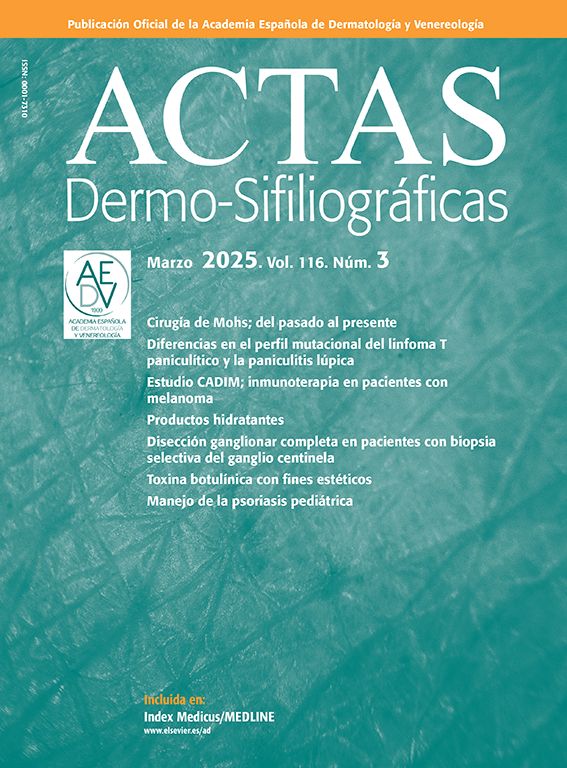


A 52-year-old women presented with a 1-year history of a slow growing yellowish and nodular plaque on the right upper eyelid (Fig. 1A). She had a recent diagnosis of bilateral carpal tunnel syndrome. Physical examination revealed a similar but more subtle lesion in the contralateral eyelid along with macroglossia and petechiae in her thighs. Using special stains, eyelid skin and tongue biopsies showed abundant amyloid deposits. Laboratory studies revealed mild normocytic anaemia, hypercreatinaemia, elevated levels of free light λ chains and a decreased serum k/λ free light chains ratio. Serum protein electrophoresis with immunofixation showed a monoclonal λ band and Bence–Jones protein was identified in the urine. Bone marrow biopsy confirmed 80% infiltration by myeloma cells. Further studies disclosed a right orbital extramedullary plasmacytoma. In one month, the eyelid plaque evolved to form an impressive mass (Fig. 1B). The patient underwent induction chemotherapy and local orbital radiotherapy with partial response. She is proposed for autologous stem cell transplantation.
In light chain amyloidosis the deposition of amyloid can occur in any organ including the skin. Clinicians, including dermatologists, face diagnostic challenges due to lack of awareness and clinical heterogeneity of this rare and serious condition.


